The Art (and Science) of Microscopes that Expand the Properties of Light We Can Perceive

“I still haven’t taken my favorite image yet,” remarks Michael Shribak, a Senior Scientist at the Marine Biological Laboratory, as he sits in his lab and discusses the latest photomicrograph of his to place within the top ten of a competition. The image, a larval sea anemone, comes from the lab of Karen Echeverri.
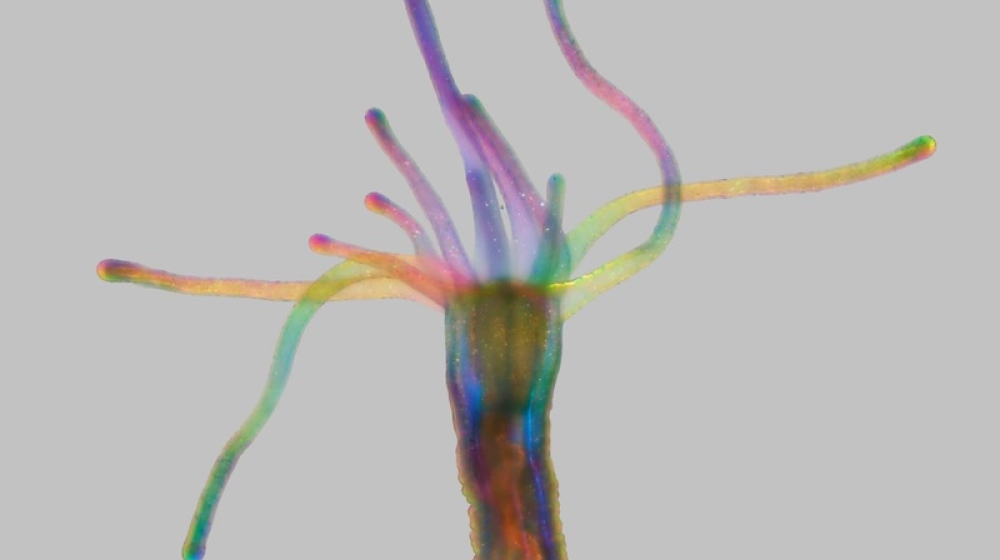
A larval sea anemone (Nematostella vectensis). Spanning just 3 millimeters across, the picture was taken using an Olympus IX83 microscope paired with a DP74 color camera and a novel label-free rainbow microscopy technique—also known as polychromatic polarizing microscopy—developed at the Marine Biological Laboratory. This patented technology (US Patent 9,625,369) is now available from the MBL for broader scientific and industrial use. Imaged by Michael Shribak and Karen Echeverri from the MBL’s Bell Center.
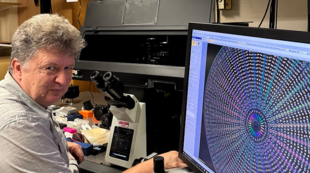
Michael Shribak with Olympus IX83 microscope equipped his polychromatic polarization module. Image credit: Michael Shribak
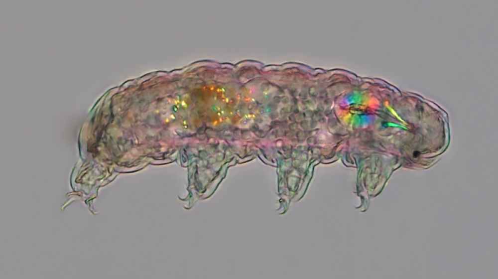
Tardigrade viewed through a Polychromatic Polarization Microscope (PPM). Imaged by Michael Shribak.
Shribak begins enlarging images of diatoms, tardigrades, and sea urchins, each containing colorful details that are usually invisible to the human eye. Humans typically perceive two properties of light: intensity (brightness) and wavelengths (color). The microscope Shribak developed and patented at the MBL in 2009, the polychromatic polarization microscope, allows him to visualize and record another property of light: Polarization.
All of Shribak’s many hued images are free of dyes and stains; their colors arise from the physics of light itself. By making the light beam of his microscope pass through a polarizing filter, similar to the filter used in a computer screen, Shribak is able to take images and videos that show the polarization of light: The direction light vibrates in as it hits a specimen.
Because his technique requires no stains or dyes that may alter a specimen’s appearance or behavior, Shribak can image live organisms in real time, capturing mesmerizing scenes of their movements under the microscope.
Today, 30 polychromatic polarization microscopes (PPM) operating in transmitted and reflected light exist worldwide. The technique has helped improve cancer detection, arthritis screening, and the identification of healthier eggs for IVF, while also finding successful applications in geology and materials science.
“PPM is a remarkable technique," explains Kazem Edmond, a senior research associate at ExxonMobil, "it should be the new standard for polarized light imaging, and it really needs to be licensed by a major vendor such as Evident Microscopy or ZEISS Microscopy.”
Edmond first began using PPM to study carbon-based materials like petroleum coke. Shribak and Edmond collaboratively developed a reflected-light version of PPM in order to obtain clear, informative images of petroleum coke microstructure features that can significantly influence its properties and behavior.
But for Shribak, the technology offers more than scientific and biomedical applications. It also provides a way to bring art and science together. “Our eyes are the best cameras, and I want to encourage people to really look,” he says.
Take a look at all the top ten winners of the BioTechniques 2026 Image Competition here.