

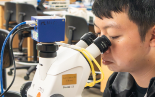


We gather 1,500 scientists and students from around the world each year.
63 Nobel Prize winners have ties to the MBL.
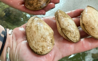
MBL scientists have studied more than 200 different kinds of marine organisms.
The MBL hosts over 200 high school students every year.