CMF Equipment
The Central Microscopy Facility supply year round and summer investigators as well as courses with instrumentation, some owned by MBL and some on loan, so please read through this guide to familiarize yourself with the Facility. Feel free to contact the Facility personnel for more information. Some of the CMF rooms are on the MBL One Card access system. You will need to get CMF authorization for your card to work on those CMF doors.
Scheduling Time and Trainings:
We use an on-line system by Stratocore for scheduling time and for setting up training sessions on individual microscopy systems. See this page for instructions.
Contact Louis Kerr at Marine Biological Laboratory, Woods Hole, Ma, 02543, Phone: (508) 289-7273, Email: lkerr@mbl.edu for more information.
Electron Microscopy

| Microscope Name: | TEM JEOL JEM-200CX |
| General Overview: | |
| 80-200KV TEM equipped with: - Low magnification mode (+/- 60 degrees tilt and 300 degree rotation) - Standard specimen cartridge and goniometer specimen cartridge - AMT digital side mount and bottom mount cameras |
|
| Commercial or Pre-commercial: | Commercial |
| Which Facility: | CMF |
| Imaging Mode: | Electron |
| Detector: | Camera |
| Location - Building and Room: | Lillie 208 |
| Contact person: | Louie Kerr |
| Hourly Rate for Use: | |
| Internal: | $36.75/hr |
| Nonprofit: | $42/hr |
| For-profit: | $73.50/hr |
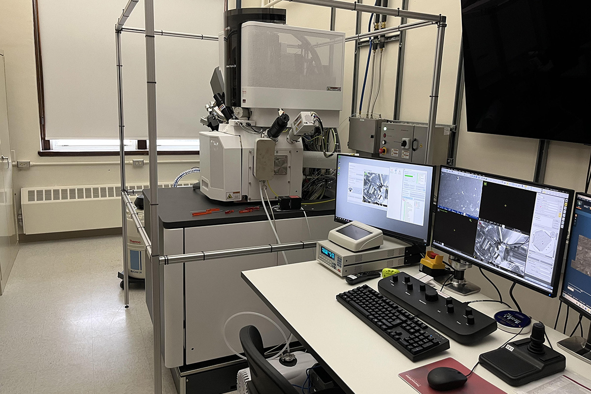
| Microscope Name: | ThermoFisher Helios 5 Hydra PFIB-SEM with Cryo and EDS/EBSD |
| General Overview: | |
| A dual-beam high-resolution scanning electron microscope with ion beam milling capability. It is equipped with multiple detectors and four different ions and can perform conventional cross-section FIB-SEM as well as spin-milling to collect larger fields of view (<115 microns) at up to 5 x 5 x 5 nm resolution. It is also equipped with cryo-imaging capabilities and EDS (Energy Dispersion Spectroscopy) and EBSD (Electron Backscatter Detection). | |
| Commercial or Pre-commercial: | Commercial |
| Which Facility: | CMF |
| Imaging Mode: | Electron |
| Features: | |
| - Plasma milling capabilities using Xenon, Argon, Nitrogen or Oxygen - Gas injection system (GIS) for deposition - ThermoFisher cryo stage with Leica transfer system and prep station - Oxford Instruments AZtec EDS/EBSD microanalysis system - ThermoFisher Auto Slice & View software for FIB-SEM acquisition - ThermoFisher MAPS software for array tomography and tile-scanning | |
| Accelerating Voltage: | - 0.5-30 kV accelerating voltage, 1.5pA-2.5uA current range |
| Temperature Control: | Yes (cryo only) |
| Additional details: | ThermoFisher Helios 5 Hydra PFIB_6357 |
| Location - Building and Room: | Lillie 207 |
| Hourly Rate for Use: | |
| Internal: | $36.75/hr for SEM use; additional $24.15/hr for use of cryo, EDS/EBSD or $50/hr for PFIB |
| Nonprofit: | $42.00/hr for SEM use; additional $28.35/hr for use of cryo, EDS/EBSD or $57.50/hr for PFIB |
| For-profit: | $73.50/hr for SEM use; additional $48.30/hr for use of cryo, EDS/EBSD or $100/hr for PFIB |
Light Microscopy
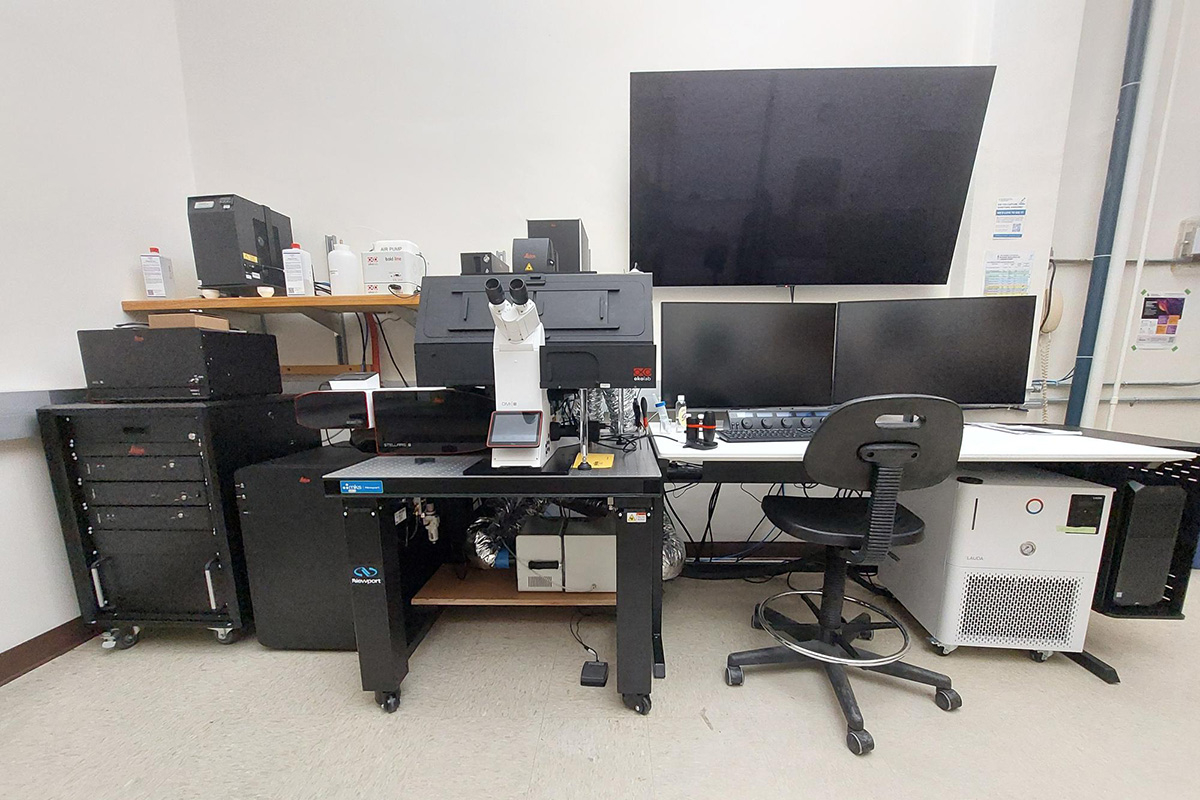
| Microscope Name: | Leica Stellaris 8 STED |
| General Overview: | |
| The Leica Stellaris is a laser-scanning confocal microscope equipped with a white light laser and tunable emission filters for maximum flexibility in excitation/emission wavelength selection. It has a environmental control chamber and features an optional z-galvo stage for rapid, smooth acquisition of 3D volumes. The Stellaris is also equipped with stimulated emission depletion (STED) super-resolution and fluorescence lifetime imaging (FLIM) capabilities. | |
| Commercial or Pre-commercial: | Commercial |
| Which Facility: | CMF |
| Imaging Mode: | Optical |
| If Optical: | Confocal, STED, FLIM |
| Microscope Stand: | Inverted |
| Features: | |
| - Leica Dmi8 microscope body with conventional and z-galvo motorized stages - Cage incubator for environmental control - 405 laser and white light laser for excitation wavelength selection - tunable emission wavelength selection - three STED lasers for super-resolution imaging - Leica FALCON FLIM analysis software |
|
| Laser Wavelengths: | 405, white light laser (450-770), STED (589, 660, 775) |
| Excitation/Emission Filters: | Tunable |
| Detector: | HyD PMTs |
| Imaging lenses: 10x/0.4 dry,20x/0.75 dry, 40x/1.1 W, 86x/1.2 W, 100x/1.4 oil (others available) |
|
| Microscope Control software: | Leica LAS X |
| Temperature Control: | Yes |
| CO2 Control: | Yes |
| Humidity Control: | Yes |
| Light Tight: | Yes |
| Sample mounting requirements/constraints: Conventional sample mounting |
|
| Location - Building and Room: | Lillie 209 |
| Hourly Rate for Use: | |
| Internal: | $34.65/hr |
| Nonprofit: | $39.90/hr |
| For-profit: | $69.30/hr |

| Microscope Name: | Zeiss LSM 780 |
| General Overview: | |
| The Zeiss LSM 780 is a multi-purpose, visible light confocal laser scanning microscope. It is equipped with a 34 channel spectral array with laser lines at 405nm, 458nm, 488nm, 514nm, 561nm, 594nm and 633nm. The LSM 780 is configured on an inverted Observer Z1 microscope with a motorized stage along with a variety of objectives. | |
| Commercial or Pre-commercial: | Commercial |
| Which Facility: | CMF |
| Imaging Mode: | Optical |
| If Optical: | Widefield, Confocal |
| Microscope Stand: | Inverted |
| Features: | Capability of spectral imaging/linear unmixing |
| Laser Wavelengths: | 405 nm, 458 nm, 488 nm, 561 nm, 594 nm, 640 nm |
| Excitation/Emission Filters: | DAPI (Zeiss 49) ; GFP (Zeiss 38HE) ; DsRed (Zeiss 43HE) |
| Detector: | PMT |
| Imaging lenses: | 5x ; 10x ; 20x ; 25xOil ; 100xOil (more lenses available) |
| Microscope Control software: | ZenBlack2.3 |
| Temperature Control: | No |
| CO2 Control: | No |
| Humidity Control: | No |
| Light Tight: | No |
| Sample mounting requirements/constraints: Conventional sample mounting |
|
| Additional details: | Zeiss LSM 710 780_small.pdf |
| Location - Building and Room: | Lillie 212 |
| Contact person: | Louie Kerr, Carsten Wolff |
| Hourly Rate for Use: | |
| Internal: | $34.65/hr |
| Nonprofit: | $39.90/hr |
| For-profit: | $69.30/hr |

| Microscope Name: | Sutter MOM 2P System |
| General Overview: | |
| The Movable Objective Microscope (MOM) from Sutter is a two-photon microscope capable of imaging deep within living specimens when combined with an appropriate laser. It provides 3-dimensional objective movement and rotation allowing the specimen to remain horizontal and stationary. | |
| Commercial or Pre-commercial: | Commercial |
| Which Facility: | CMF |
| Imaging Mode: | Optical |
| If Optical: | Confocal, Multi-photon |
| Microscope Stand: | adjustable position (upright or inverted or in-between) |
| Features: | Objective rotates about optical axis for imaging; customizable open platform design |
| Laser Wavelengths: | Coherent Chameleon Ultra2 laser offers a tuning range from 680nm to 1080nm |
| Detector: | PMT |
| Imaging lenses: | 20xW, 0.8NA ; other lenses available |
| Microscope Control software: | MScan3.0 |
| Temperature Control: | No |
| CO2 Control: | No |
| Humidity Control: | No |
| Light Tight: | No |
| Sample mounting requirements/constraints: | Conventional sample mounting as well as custom solutions |
| Additional details: | COHR_Chameleon_ultra2_laser.pdf Sutter_MOM_2Pr.pdf |
| Location - Building and Room: | Rowe 203 |
| Contact person: | Louie Kerr, Carsten Wolff |
| Hourly Rate for Use: | |
| Internal: | $34.65/hr |
| Nonprofit: | $39.90/hr |
| For-profit: | $69.30/hr |

| Microscope Name: | Zeiss Axio Observer Spinning Disc |
| General Overview: | |
| The Zeiss Cell Observer Spinning Disk confocal system has a Photometrics Evolve 512×512 camera. It is configured on an inverted Observer Z1 microscope with a motorized stage and a complete incubator chamber system. | |
| Commercial or Pre-commercial: | Commercial |
| Which Facility: | CMF |
| Imaging Mode: | Optical |
| If Optical: | Widefield, Confocal |
| Microscope Stand: | Inverted |
| Features: | Electrophysiology equipment and FRAP module ; Evolve 512 Delta EMCCD Camera |
| Laser Wavelengths: | 488 nm, 561 nm, 635 nm |
| Excitation/Emission Filters: | DAPI (Zeiss 49) ; GFP (Zeiss 38HE) ; DsRed (Zeiss 43HE) ; BS C/G/DR |
| Detector: | Camera |
| Imaging lenses: | 10x ; 20x ; 40xOil ; 40xW ; 100xOil ; 150xGly |
| Microscope Control software: | ZenBlack2 |
| Temperature Control: | Yes |
| CO2 Control: | Yes |
| Humidity Control: | Yes |
| Light Tight: | No |
| Sample mounting requirements/constraints: | Conventional sample mounting |
| Additional details: | Evolve512-Datasheet.pdf |
| Location - Building and Room: | Loeb G63 |
| Contact person: | Louie Kerr, Carsten Wolff |
| Hourly Rate for Use: | |
| Internal: | $25.20/hr |
| Nonprofit: | $29.40/hr |
| For-profit: | $50.40/hr |

| Microscope Name: | Zeiss Lightsheet7 |
| General Overview: | |
| The Zeiss Lightsheet 7 is a stand-alone SPIM (Selective Plane Illumination Microscope) capable of imaging large, living specimens at much greater speeds than more traditional confocal technology. The fully motorized sample chamber translates in X,Y and Z as well as 360° rotation, which allows optical sectioning from virtually any angle within the specimen. The chamber is equipped for live imaging with control over temperature and CO². | |
| Commercial or Pre-commercial: | Commercial |
| Which Facility: | CMF |
| Imaging Mode: | Optical |
| If Optical: | Widefield, Lightsheet |
| Microscope Stand: | Sample get introduced vertically into the microscope chamber. |
| Features: | One microscope chamber is larger and especially designed for holding media used for sample tissue clearing ; Camera: PCO edge 4.2 sCMOS |
| Laser Wavelengths: | 405 nm, 488 nm, 561 nm, 647 nm |
| Excitation/Emission Filters: | DAPI (Zeiss 49) ; GFP (Zeiss 38HE) ; DsRed (Zeiss 43HE) ; Cy5 (Zeiss 50) |
| Detector: | Camera |
| Imaging lenses: | Illumination: 5x Multi Immersion ; 10x Multi Immersion Detection: 5x Multi Immersion ; 10x (Zeiss loan) ; 20x Multi Immersion |
| Microscope Control software: | ZenBlack |
| Temperature Control: | Yes |
| CO2 Control: | Yes |
| Humidity Control: | Yes |
| Light Tight: | Yes |
| Sample mounting requirements/constraints: | Microscope chamber is virtually built around your sample, with its holder designed to best support your experiment's purpose. Sample holder interface is open both to custom design and machining and 3D printing of your own sample holder. |
| Additional details: | Lightsheet-7_info.pdf pco.edge_42_A_data_sheet.pdf |
| How to list instrument for inclusion in papers, reports and grant proposals: | The ZEISS Lightsheet 7 was supported by the Howard Hughes Medical Institute. |
| Location - Building and Room: | Loeb G58 |
| Contact person: | Louie Kerr, Carsten Wolff |
| Hourly Rate for Use: | |
| Internal: | $36.75/hr |
| Nonprofit: | $42.00/hr |
| For-profit: | $73.50/hr |

| Microscope Name: | diSPIM |
| General Overview: | |
| This microscope offers low photo-toxicity and large specimen handling of dual inverted selective plane illumination microscopy (diSPIM) with the power and flexibility of a live-cell microscope system. This system is suitable for imaging samples in aqueous media. | |
| Commercial or Pre-commercial: | Pre-Commercial |
| Which Facility: | CMF, Imaging Innovation Initiative |
| Imaging Mode: | Optical |
| If Optical: | Lightsheet |
| Microscope Stand: | Custom designed for upright applications |
| Features: | Suitable for imaging large samples at high volumetric speed. |
| Laser Wavelengths | 405 nm, 488 nm, 561 nm, 640 nm |
| Excitation/Emission Filters: | Quad band filter for 405/488/561/640 lasers. |
| Detector: | Camera |
| Imaging lenses: | 14x, 0.4 NA Multi-immersion lenses; 23x, 0.7 NA Multi-immersion lenses |
| Microscope Control software: | MicroManager |
| Temperature Control: | Yes |
| CO2 Control: | No |
| Humidity Control: | No |
| Light Tight: | No |
| Sample mounting requirements/constraints: | Conventional sample mounting |
| Additional details: | diSPIM_Kumar_etal2014_NatureProtocols.pdf |
| Location - Building and Room | Lillie 215 |
| Contact person: | Louie Kerr, Carsten Wolff |
| Hourly Rate for Use: | |
| Internal: | $34.65/hr |
| Nonprofit: | $39.90/hr |
| For-profit: | $69.30/hr |

| Microscope Name: | Zeiss Axio Imager.M2 - Manual Stage |
| General Overview: | |
| Upright compound microscope set up for transmitted light techniques such as brightfield, phase and DIC, and also set up for epi-fluorescence with a X-Cite light source. It includes a a motorized Z-drive and Zeiss AxioCams (color and b/w). | |
| Commercial or Pre-commercial: | Commercial |
| Which Facility: | CMF |
| Imaging Mode: | Optical |
| If Optical: | Widefield |
| Microscope Stand: | Upright |
| Features: | Epifluorescence ; Cameras: Zeiss AxioCam 503 (b/w) ; Zeiss AxioCam 305 (color) |
| Excitation/Emission Filters: | DAPI (Zeiss 49) ; AF488 (Zeiss 09) ; GFP (Zeiss 38HE) ; DsRed (Zeiss 43HE) ; Cy5 (Zeiss 50) |
| Detector: | Camera |
| Imaging lenses: | 5x ; 10x ; 20x ; 63xOil ; 63xW ; 100xOil (more lenses available) |
| Microscope Control software: | ZenBlue3.1 |
| Temperature Control: | No |
| CO2 Control: | No |
| Humidity Control: | No |
| Light Tight: | No |
| Sample mounting requirements/constraints: | Conventional sample mounting |
| Additional details: | AxioImagerM2.pdf |
| Location - Building and Room: | Lillie 209 |
| Contact person: | Louie Kerr, Carsten Wolff |
| Hourly Rate for Use: | |
| Internal: | $12.60/hr |
| Nonprofit: | $14.70/hr |
| For-profit: | $25.20/hr |

| Microscope Name: | Zeiss Axio Observer |
| General Overview: | |
| The Zeiss Axio Observer is set up for transmitted light techniques such as brightfield, phase and DIC and epi-fluorescence with a Colibri LED illuminator and mercury halide lamp. It includes a motorized stage and a Zeiss AxioCam 702. | |
| Commercial or Pre-commercial: | Commercial |
| Which Facility: | CMF |
| Imaging Mode: | Optical |
| If Optical: | Widefield |
| Microscope Stand: | Inverted |
| Excitation/Emission Filters: | DAPI (Zeiss 49) ; GFP (Zeiss 38HE) ; DsRed (Zeiss 43HE) |
| Detector: | Camera |
| Imaging lenses: | 2.5x ; 5x ; 10x ; 20x ; 40xOil ; 63xOil / Optovar 1x ; 1.6x |
| Microscope Control software: | ZenBlue3.4 |
| Temperature Control: | Yes |
| CO2 Control: | Yes |
| Humidity Control: | Yes |
| Light Tight: | No |
| Sample mounting requirements/constraints: | Conventional sample mounting |
| Location - Building and Room: | Loeb G61 |
| Contact person: | Louie Kerr, Carsten Wolff |
| Hourly Rate for Use: | |
| Internal: | $12.60/hr |
| Nonprofit: | $14.70/hr |
| For-profit: | $25.20/hr |

| Microscope Name: | Zeiss Axio Zoom V16 Stereo Microscope |
| General Overview: | |
| The Zeiss Axio Zoom V16 is a stereomicroscope that features a very high numerical aperture over a large field of view. This allows for bright fluorescence imaging of samples from 7x-112x magnification. It is a completely motorized stereomicroscope with automated Z and stage for 3D imaging as well as tiling that combine the highest magnification with the largest field of view. | |
| Commercial or Pre-commercial: | Commercial |
| Which Facility: | CMF |
| Imaging Mode: | Optical |
| If Optical: | Widefield |
| Microscope Stand: | Upright |
| Features: | Range of magnification : 7x-112x Equipped with Apotome2 Cameras: Zeiss AxioCam 712 (b/w) ; Zeiss AxioCam 512 (color) |
| Excitation/Emission Filters: | DAPI (Zeiss 49) ; GFP (Zeiss 38HE) ; DsRed (Zeiss 43HE), Cy5 (Zeiss50) |
| Detector: | Camera |
| Imaging lenses: | |
| Microscope Control software: | ZenBlue3.4 |
| Temperature Control: | No |
| CO2 Control: | No |
| Humidity Control: | No |
| Light Tight: | No |
| Sample mounting requirements/constraints: | Conventional sample mounting |
| Additional details: | Zeiss Axio-Zoom-V16.pdf |
| Location - Building and Room | Lillie 210 |
| Contact person: | Louie Kerr, Carsten Wolff |
| Hourly Rate for Use: | |
| Internal: | $12.60/hr |
| Nonprofit: | $14.70/hr |
| For-profit: | $25.20/hr |

| Microscope Name: | Zeiss Axio Zoom V12 Stereo Microscope |
| General Overview: | |
| The Discovery V12 is a stereo dissecting microscope set up for transmitted light techniques. It includes a VisiLED base. | |
| Commercial or Pre-commercial: | Commercial |
| Which Facility: | CMF |
| Imaging Mode: | Optical |
| If Optical: | Widefield |
| Microscope Stand: | Upright |
| Features: | Equipped with a VisiLED base Camera: Zeiss AxioCam MRc |
| Detector: | Camera |
| Microscope Control software: | AxioVision |
| Temperature Control: | No |
| CO2 Control: | No |
| Humidity Control: | No |
| Light Tight: | No |
| Sample mounting requirements/constraints: | Conventional sample mounting |
| Location - Building and Room | Lillie 210 |
| Contact person: | Louie Kerr, Carsten Wolff |
| Hourly Rate for Use: | |
| Internal: | $12.60/hr |
| Nonprofit: | $14.70/hr |
| For-profit: | $25.20/hr |
Other Equipment
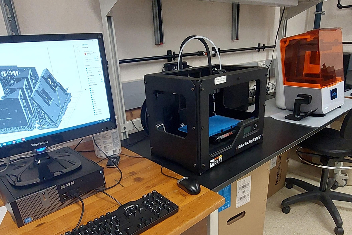
| Equipment Name: | 3D printer Formlabs Form 3B+ |
| General Overview: | |
| Formlabs Form 3B+ is an industrial-quality desktop 3D printer that can produce functional, high-quality prototypes and end-use parts in record time. The Form 3+ offers reliable print quality and accuracy, easy setup and maintenance, and a wide range of high-performance materials. | |
| Commercial or Re-commercial: | Commercial |
| Which Facility: | CMF |
| Features: | - Printing resolution 25-300μm (resin layer thickness); - Build volume 14.5 × 14.5 × 18.5 cm (5.7 × 5.7 × 7.3 in); - Printing resins available in: black, grey, clear, clear bio-compatible , flexible80A / more available by request; - FormWash and FormCure for post-processing; - Workstation with essential software; |
| Additional details: | Formlabs_3B_manual.pdf Formlabs_3B_quick guide.pdf Formlabs_FormCure.pdf Formlabs_FormWash.pdf |
| Location - Building and Room: | Lillie 215 |
| Contact person: | Louie Kerr, Carsten Wolff |
| Hourly Rate for Use: | |
| Internal: | $11.00 (per 10mL resin or 5g filament) |
| Nonprofit: | $12.65 |
| For-profit: | $22.00 |

Imaris 9.5.1
Avia 11
Arivis
ZEN Blue 3.6
MATLAB
Fiji / ImageJ
Napari