Multiplex Microscopy Becomes Easier with Encoded Antibodies | The Scientist Digest
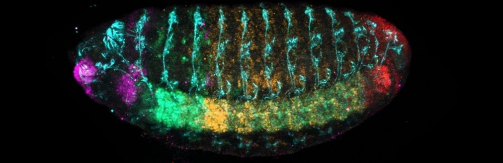
Editor’s note: This article features insights from Nipam Patel, the director of the MBL.
Nipam Patel, a developmental biologist at the University of Chicago, and his group study the development of body morphology in arthropods, specifically crustaceans in the Parhyale genus. The team focuses on the role and impact of various homeotic (Hox)genes and their proteins, which encode information to guide the animal’s body plan and the role and development of various limbs.
In Patel’s team’s crustaceans, the researchers have seen these genes have unique functions for appendage patterns that are distinct from the often-studied fly. “It explains how crustaceans can generate so much diversity in their appendage types,” he said. The team explores the role of these genes in body segmentation with a combination of genetic models and fluorescent microscopy.
Because Hox proteins work together to give rise to an animal’s body plan, Patel and his team often want to visualize multiple targets simultaneously in microscopy. However, because most monoclonal antibodies come from mice, this introduces challenges for multiplexing since all the antibodies have the same species origin.
Read more of the article here.
Source: Multiplex Microscopy Becomes Easier with Encoded Antibodies | The Scientist Digest