New MBL Microscope Combines Two Powerful Imaging Techniques

The Marine Biological Laboratory has purchased a new, advanced confocal microscope with a grant from the National Institutes of Health awarded to Associate Scientist Michael Shribak.
Shribak, in collaboration with Kazuhiro Maeshima of Japan's National Institute of Genetics (NIG) and MBL's Tomomi Tani, will use the microscope to study heterochromatin (a condensed form of DNA) in living tissues. Previously, the team's work required two separate imaging steps: obtaining a dry mass map of the samples at the MBL using a technique called quantitative orientation-independent differential interference contrast (OI-DIC) microscopy, followed by confocal microscopy at NIG in Japan. The need to perform imaging in two widely separated locales obviously complicated the experimental process as well as incurring increased expense.
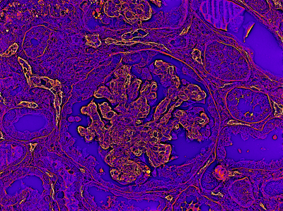 OI-DIC image of immunohistochemistry for complement C4d component in kidney allograft. Credit: Michael Shribak (MBL) and Arvydas Laurinavičius (Vilnius University)
The new microscope, an Olympus FV3000, combines confocal laser scanning with OI-DIC imaging capabilities to produce quantitative, high-resolution, thin-section images. Shribak's team also recently showed that OI-DIC microscopy, which Shribak invented, can exceed the Abbe limit (the resolution limit of light microscopes), which will facilitate other research efforts at MBL. Unlike confocal microscopy, which uses a focused laser beam that can damage live organisms, OI-DIC employs a beam from a conventional halogen or LED lamp, which is safe for living tissues. The combination of these two powerful microscopy techniques in a single instrument allows for a wide range of high-resolution and 3D imaging experiments to be performed at MBL.
After receiving the new microscope in spring 2019, Shribak and his team will begin using it for new experiments in the summer.