Embryology tries out new tools for probing biodiversity

The second floor of MBL’s Loeb Laboratory will be populated by a menagerie of organisms this summer, ranging from the well-studied, such as flies and frogs, to marine and other organisms whose biology remains to be fully grasped. Students in the Embryology course are in this lab space day and night, devising experiments and exploring the latest research "tools" that are loaned to the MBL courses by hundreds of vendors.
“Because we look at so many diverse animals, students have the rare chance to work with less-studied models and make comparisons across species,” says course co-director Richard Behringer of the MD Anderson Cancer Center, University of Texas.

The archetypal “Woods Hole experiment," as Behringer describes it, is a project so novel that the students—who are graduate students, postdoctoral fellows, or senior researchers from around the world—would shy away from the challenge in any other context. “This course gives them the courage to step outside their comfort zones and break self-imposed barriers,” he says.
On a recent afternoon, student Mike Abrams of Caltech is testing the waters of sea urchin development using a newly minted system that displays microscope images and data on an iPad screen. Abrams is interested in the repercussions of injecting sea-urchin embryonic cells into a starfish embryo. "My work is now much more mobile," he says; if he has a question, he can detach the iPad and go find help. “I don’t have to wait for the TA to come to me!” The iPad-scopes are linked, "so I can instantaneously see files from classmates’ iPad-microscopes without leaving my own lab bench. Most importantly, there’s no need to refocus the image between each new set of eyes," he says. Behringer hopes to obtain six more iPad-enabled microscopes to equip every student in the class with one.
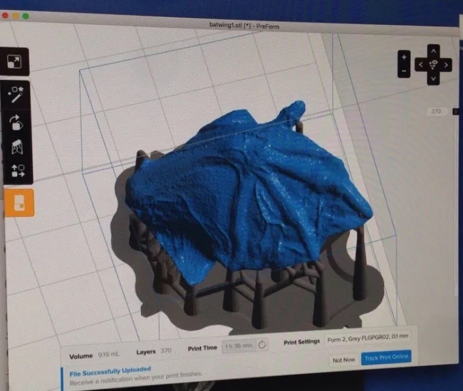
The iPad-scopes are not the only recent addition to the Embryology course toolkit. Faculty member Jon Henry of the University of Illinois borrowed a 3D printer for the lab as well. As part of a separate project, Abrams has teamed up with Kate Woronowicz of University of California-San Francisco to collect a “stack” of images to print an object reminiscent of a spaceship—which is actually a replica of the sea-urchin larval skeleton. “In the future, we could use the printer to create physical representations of organisms with specific genetic mutations,” Woronowicz says.
Behringer emphasizes the technology’s educational potential. “If you give a student a three-dimensional object to hold, they’ll understand it better than an image in a textbook,” he says. While the 3D printer and iPad-associated microscopes are still in the nascent stages of scientific application, in a setting like the MBL Discovery Courses, the possibilities are manifold.