Microscope Allows Gentle, Continuous Imaging of Light-Sensitive Corals

WOODS HOLE, Mass. – Corals are “part animal, part plant, and part rock - and difficult to figure out, despite being studied for centuries,” says Philippe Laissue of University of Essex, a Whitman Scientist at the Marine Biological Laboratory. Many corals are sensitive to bright light, so capturing their dynamics with traditional microscopes is a challenge.
To work around their photosensitivity, Laissue developed a custom light-sheet microscope (the L-SPI) that allows gentle, non-invasive observation of corals and their polyps in detail over eight continuous hours, at high resolution. He and his colleagues, including MBL Associate Scientist and coral biologist Loretta Roberson, published their findings this week in Scientific Reports.
Below is a video explainer of the research:
Coral reefs, made up of millions of tiny units called polyps, are extremely important ecosystems, both for marine life and for humans. They harbor thousands of marine species, providing food and economic support for hundreds of millions of people. They also protect coasts from waves and floods, and hold great potential for pharmaceutical and biotechnological discovery.
But more than half of the world’s coral reefs are in severe decline. Climate change and other human influences are gravely threatening their survival. As ocean temperatures rise, coral bleaching is afflicting reefs worldwide. In coral bleaching, corals expel their symbiotic algae and become more susceptible to death.

“The L-SPI opens a window on the interactions and relationship between the coral host, the symbiotic algae living in their tissues, and the calcium carbonate skeleton they build in real time,” Roberson says. “We can now track the fate of the algae during [coral] bleaching as well as during initiation of the symbiosis.”
Roberson is also using Laissue’s imaging technology to measure damage to corals from “bioeroders” – biological agents like algae and sponges that break down a coral’s skeleton, a problem exacerbated by ocean acidification and increasing water temperatures.
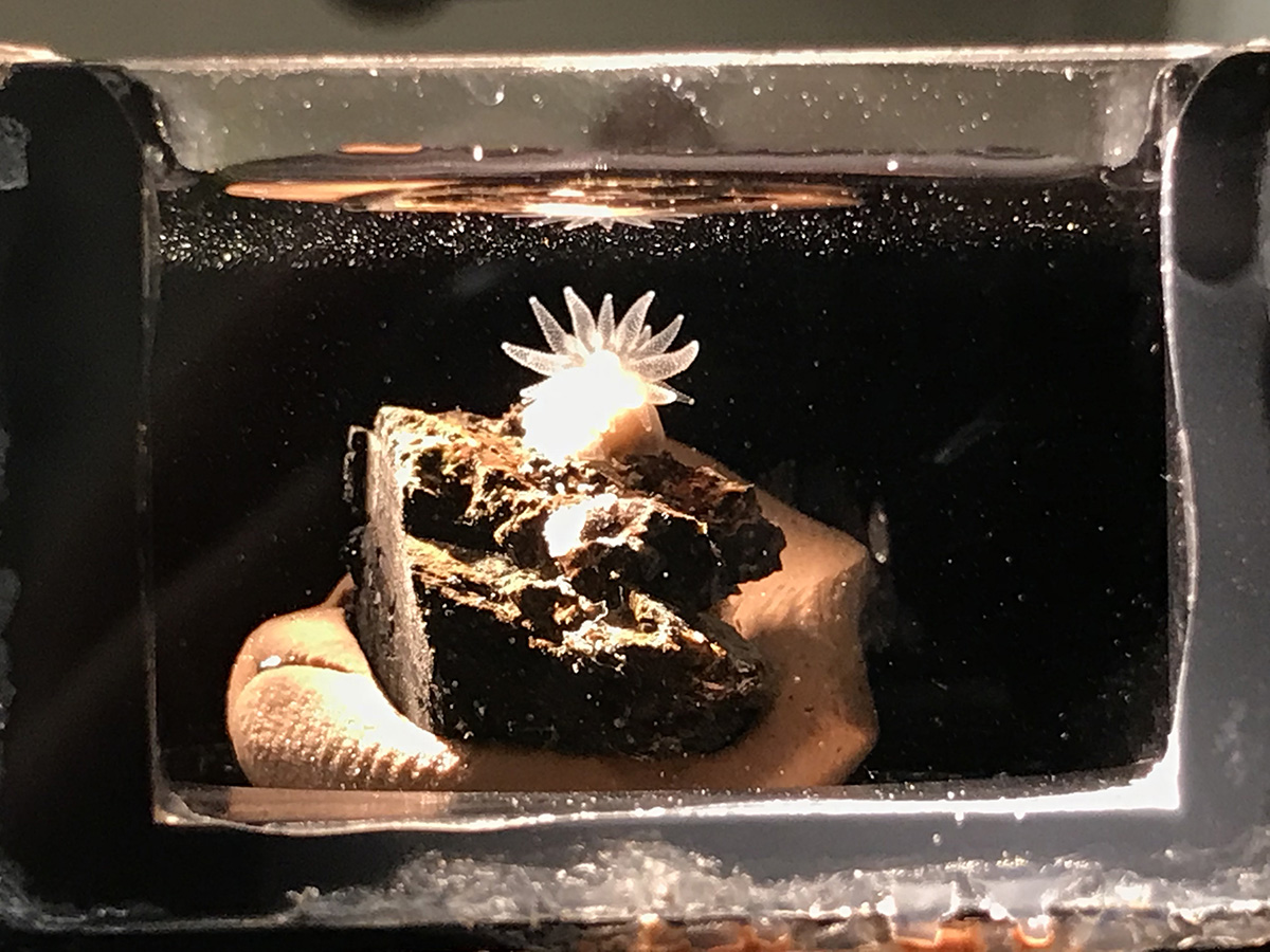 A coral polyp (Astrangia) in the L-SPI sample chamber with tentacles fully extended. Credit: Loretta Roberson
Laissue has been coming regularly to the MBL for research since obtaining a Whitman Center Fellowship in 2017. He is continuing to collaborate with Roberson and MBL Fellow Amy Gladfelter, and to develop the L-SPI microscope with MBL-CZI Imaging Scientist Abhishek Kumar and MBL Fellow Hari Shroff.
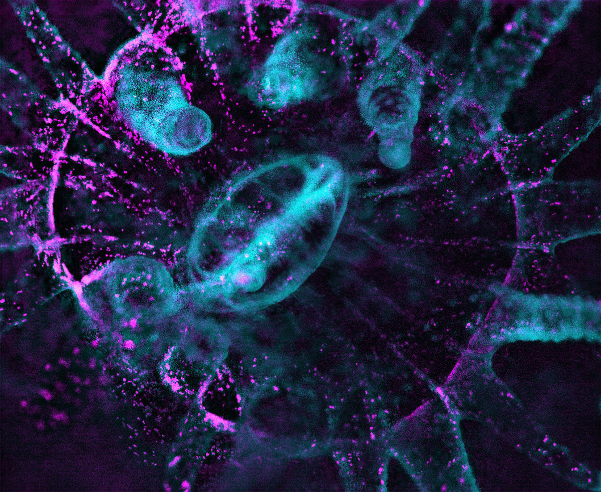 Darkfield imaging is used to reveal the non-autofluorescent coral tissue in the Northern Star coral (Astrangia poculata), a species with no intrinsic fluorescence. Non-fluorescent tissue (cyan) and algae (magenta) are combined in the image. Credit: Philippe Laissue, Scientific Reports, 2020
A video of a coral polyp emerging that Laissue imaged with the L-SPI microscope won first place in the 2019 Nikon Small World in Motion. See the video here.
Citation: Laissue, P.P., Roberson, L., Gu, Y. et al. (2020) Long-term imaging of the photosensitive, reef-building coral Acropora muricata using light-sheet illumination. Scientific Reports, DOI: 10.1038/s41598-020-67144-w
—###—
The Marine Biological Laboratory (MBL) is dedicated to scientific discovery – exploring fundamental biology, understanding marine biodiversity and the environment, and informing the human condition through research and education. Founded in Woods Hole, Massachusetts in 1888, the MBL is a private, nonprofit institution and an affiliate of the University of Chicago.