Snapshots of Life: Neurons in a New Light | NIH Director's Blog

Francis Collins, director of the National Institutes of Health, blogs about an innovative polarized light microscope recently developed by MBL Associate Scientist Michael Shribak. The microscope can generate detailed images of cells and tissues -- including neurons as in the image at right -- without perturbing them with stains or dyes. The microscope can also be used to study a range of disorders, from cancer to blood cell disorders such as sickle cell disease and malaria.

Birds do it, bees do it, and even educated fleas do it. No, not fall in love, as the late Ella Fitzgerald so famously sang. Birds and insects can see polarized light—that is, light waves transmitted in a single directional plane—in ways that provides them with a far more colorful and detailed view of the world than is possible with the human eye.
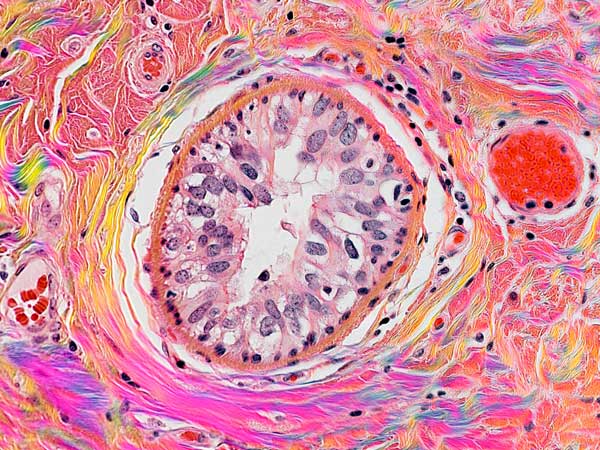 Polarization polychromatic image of birefringent collagen fibers in H&E-stained breast cancer tissue. Credit: Michael Shribak and Richard Levenson
Still, thanks to innovations in microscope technology, scientists have been able to tap into the power of polarized light vision to explore the inner workings of many complex biological systems, including the brain. In this image, researchers used a recently developed polarized light microscope to trace the spatial orientation of neurons in a thin section of the mouse midbrain. Neurons that stretch horizontally appear green, while those oriented at a 45-degree angle are pinkish-red and those at 225 degrees are purplish-blue. What’s amazing is that these colors don’t involve staining or tagging the cells with fluorescent markers: the colors are generated strictly from the light interacting with the physical orientation of each neuron.
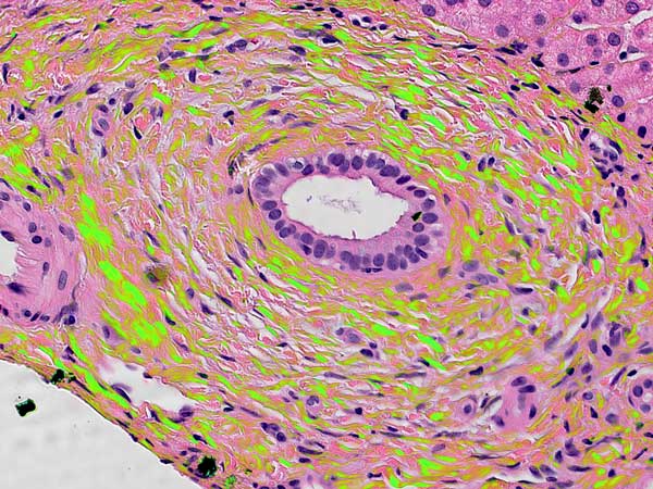 Polarization polychromatic image of birefringent collagen fibers in H&E-stained liver cancer tissue. Credit: Michael Shribak and Richard Levenson
Source: Snapshots of Life: Neurons in a New Light | NIH Director's Blog