High School Students Discover Science in the Nipam Patel Lab

By Sophia Kelly Patel Lab, Marine Biological Laboratory
This month, we were fortunate to take a break from our day-to-day lab work to host a group of 25 high-school students from the Glenbard East High School in Lombard, Illinois, and the Hockaday School in Dallas, Texas. The course was part of the MBL's new High School Science Discovery Program.
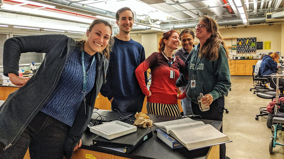 Left to right: Nipam Patel lab members Sophia Kelly, Kyle DeMarr, Heather Bruce, Nipam Patel, and Jenny McCarthy share some laughs while helping out with the high-school course!
We reinforced Nipam’s colorful lectures on development through hands-on lab training with the students. They were able to set up their own zebrafish embryo timelapse videos, discover how colors are produced in butterfly wings, and complete antibody stains on various embryos.
The students watched their zebrafish embryos develop. Also, using a fluorescence microscope, they watched the heart beating and blood moving through other zebrafish embryos tagged with green fluorescent protein (GFP Fli Line).
We also taught the students about antibody staining, which allows for detection of specific proteins throughout a sample. The students completed their own stains on fly, zebrafish, squid, and cuttlefish embryos.
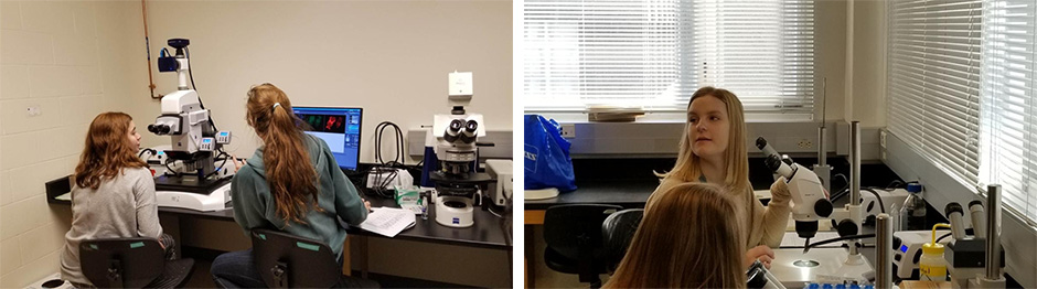 Left: Jenny McCarthy of the Patel Lab and Gabrielle Spontak of Glenbard East look at stained fly embryos under the Zeiss AxioZoom V16 microscope. Right: Megan High of Glenbard East mounts fly embryos under a dissecting microscope. Photos by Glenbard East chaperone Marisa Abrams.
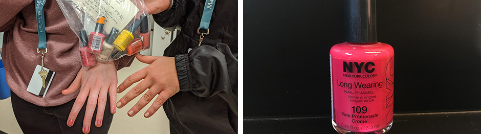
They then learned how to mount their antibody stains under dissecting and fluorescence microscopes. They were very amused at our use of NYC Pink Promenade nail polish to seal down our microscope cover slips!
Besides the fact that this nail polish is an amazing, vibrant pink, we use it because it does not fluoresce under the lasers of the confocal microscope and is the perfect consistency, as it does not spread out all over the slide. We have been having a hard time finding this Pink Promenade for sale anywhere, so Patel lab manager, Erin Jarvis, purchased a wide range of different NYC polishes. Two of the students graciously tested out these polishes for us, returning the next day with detailed notes on their consistencies, colors and dry times. (Thank you, Paige and Helena!) Who said fashion isn’t important for science?!
 Once the students had their antibody stains mounted, Nipam showed them how to use the confocal microscope and they took images for processing in ImageJ, a Java-based image processing program.
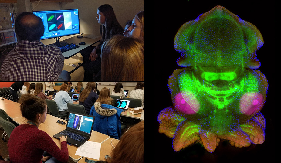 Top left: Students take images of their antibody stains on the confocal microscope with Nipam Patel. Bottom left: Students analyze and process their images in ImageJ. Photos taken by Glenbard East chaperone Marisa Abrams. Right: Squid embryo confocal image taken by Tia Hsieh (Hockaday), imaged on a Zeiss LSM 880 and processed using ImageJ.
Last, but definitely not least, the students explored the beauty of butterfly wings. Through a simple experiment with acetone and xylene, they examined different species of butterflies to understand if their wing scales exhibit structural colors, pigmentary colors, or both! They even got a close-up view of the wing scales on a microscope in our lab.
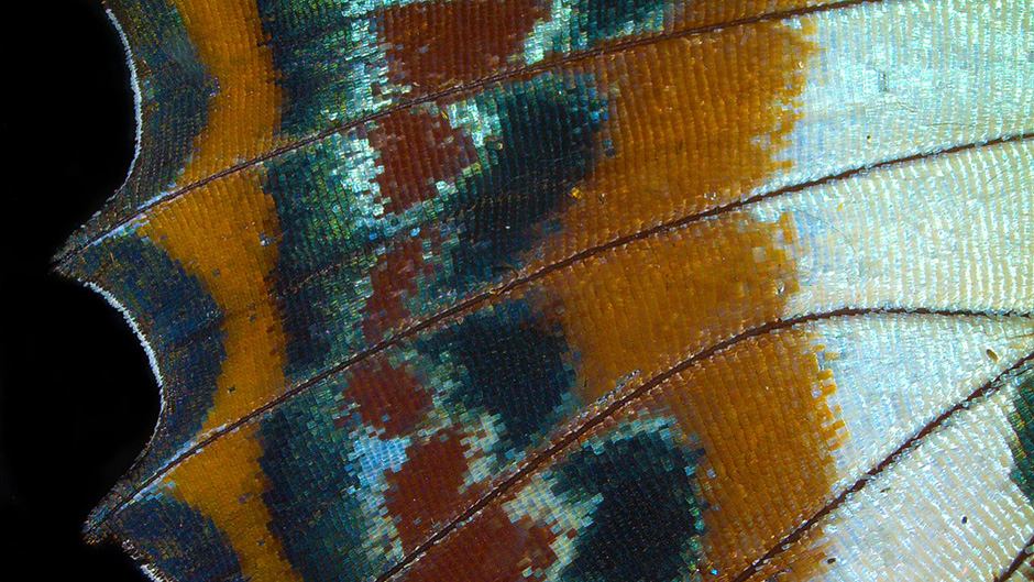 The wing of Charaxes brutus, a butterfly from the Central African Republic. Image by Daniela Vallejo (Hockaday), taken on a Zeiss Stemi 508 using an AxioCam ERc 5 and LabScope software.
We were so pleased with the students’ enthusiasm throughout the week, especially when they are dealing with the chaos of finishing up high school and applying to colleges (Good luck!). It is always a pleasure working with young, bright-eyed students, as we can only hope that spending time at the Marine Biological Laboratory will help inspire future generations of scientists to come!
Click here to learn more about MBL’s educational offering for secondary education students.