Full Name
Margherita Perillo
Title
Research Scientist

- Email:
- Phone:
- Fax:
- CV:
- ORCID ID:
0000-0003-0845-507X
M.Sc. with Honors, Biotechnology of Drugs, Università degli Studi di Napoli Federico II, 2008
B.S. Health Biotechnologies, Università degli Studi di Napoli Federico II, 2005
We are interested in the cellular and molecular mechanisms that guide organ morphogenesis. During embryogenesis, individual cells group together to build organs with complex three-dimensional structures. Disruption of the perfect balance that coordinates the many cells in the organ epithelium can cause major embryonic malformations, organ displacement and loss of function, and defects in organ regeneration in the body. How do unique cell-types contribute to organ homeostasis? What are the cues that guide the migration of individual cells to their final position in the organ? How did the first organs evolve?
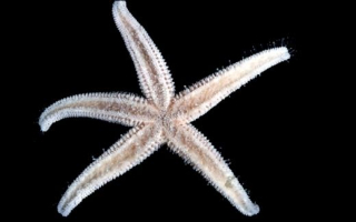
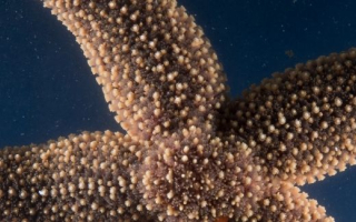
To answer these questions, we use the sea star Patiria miniata as a model system to study several aspects of organ development and evolution. Sea stars are marine animals with a close evolutionary relationship to vertebrates, and therefore can help us understand the evolution of organs. Moreover, sea star larvae are optically transparent and genetically tractable, making it a suitable system to study organogenesis in an intact organism.

The main themes of our research are:
Organogenesis: Mechanisms of epithelial tube formation. Most vital organs, including lungs, kidneys, and mammary glands, are organized into tubular shapes with the essential scope of transporting gasses, liquids, or cells. The three-dimensional architecture of these structures is fundamental to their function. What signaling pathways and cellular mechanisms drive the formation of tubular organs? Sea star larvae swim in the ocean, and their movement requires the hydro-vascular organ, a tubular structure with one opening towards the outside environment. First, we are defining how the hydro-vascular organ works and its role in the life of the tiny sea star larva. Second, we use this model to uncover 1) how individual cells organize into organs, and 2) the factors that disrupt the perfect harmony of cells that coexist in a healthy organ, as in cancer and other pathologies.
Evolutionary origins of organs. The first animals lived in the ocean, and the interactions between the embryo and the outside environment likely shaped the function and the architectures of primordial organs. How did the first organs evolve? Understanding the origin of organs requires species comparisons. Some species develop hydro-vascular organs with a diversity of shapes and unique development. To determine how this three-dimensional diversity is achieved, we take an evo-devo approach by comparing organogenesis in different echinoderm larvae, such as sea cucumbers, sea urchins and of course sea stars.

Mechanisms of collective cell migration. Collective cell behaviors ensure spatio-temporal coordination among the group of cells that results in correct tissue patterning. Primordial germ cells (PGCs), the precursors to germline stem cells, are an example of cell types that undergo coordinated collective movement to ensure correct tissue patterning. We found that in the sea star larva, the PGCs migrate as a cluster and eventually merge with the hydro-vascular organ. What are the cues that guide PGC migration to their final niche? To answer this question, we use a combination of biochemistry, live imaging and transcriptomics.


Google Scholar: https://scholar.google.com/citations?user=g9WHqL8AAAAJ&hl=it
Marc, T. K., Rodger, A., & Perillo, M. (2026). Comparative analysis of Asterias forbesi development reveals distinct mechanisms of hydro-vascular organ formation across sea stars. Discover Developmental Biology, 236(1), 4.
Clarke DN, Kane A, Perillo M, Lowe CJ, Swartz SZ. VitelloTag: a tool for high-throughput cargo delivery into oocytes. Development. 2024 Oct 15;151(20). doi: 10.1242/dev.202857. Epub 2024 Sep 14. PubMed PMID: 39171380; PubMed Central PMCID: PMC11423919.
Perillo, M.,* Alessandro T., Toscano, A., & Annunziata, R.* (2024). Larval development of Holothuria tubulosa, a new tractable system for evo-devo. Front. Ecol. Evol. doi: 10.3389/fevo.2024.1409174. *Co-corresponding authors
Perillo, M.,* Sepe, R. M., Paganos, P., Toscano, A., & Annunziata, R.* (2024). Sea cucumbers: an emerging system in evo-devo. EvoDevo, 15. *Co-corresponding authors
Perillo, M.*, Swartz, S. Z., Pieplow, C., & Wessel, G. M.* (2023). Molecular mechanisms of tubulogenesis revealed in the sea star hydro-vascular organ. Nature Communications, 14(1), 1-17. *Co-corresponding authors
Spurrell, M., Oulhen, N., Foster, S., Perillo, M., & Wessel, G. (2023). Gene regulatory divergence amongst echinoderms underlies appearance of pigment cells in sea urchin development. Developmental biology, 494, 13-25.
Oulhen, N., Pieplow, C., Perillo, M., Gregory, P., & Wessel, G. M. (2022). Optimizing CRISPR/Cas9-based gene manipulation in echinoderms. Developmental biology 490, 117-124
Perillo, M., Swartz, S. Z., & Wessel, G. M. (2022). A conserved node in the regulation of Vasa between an induced and an inherited program of primordial germ cell specification. Developmental Biology, 482, 28-33.
Swartz, S. Z., Tan, T. H., Perillo, M., Fakhri, N., Wessel, G. M., Wikramanayake, A. H., & Cheeseman, I. M. (2021). Polarized Dishevelled dissolution and reassembly drives embryonic axis specification in sea star oocytes. Current Biology, 31(24), 5633-5641.