Bats and Chameleons, Oh My! MBL Alumni Place in Nikon Photo Contest
A glowing bat and a curled chameleon, both imaged by MBL Embryology Course students, are among the top 20 winners of the 2020 Nikon Small World Photomicrography Competition, a leading forum for showcasing life as seen through a light microscope.
The chameleon, which placed 8th, was taken by 2019 Embryology course students Allan Carrillo-Baltodano and David Salamanca. “Allan and I usually work with small invertebrates, but we were eager to learn, not matter what!” Salamanca says.
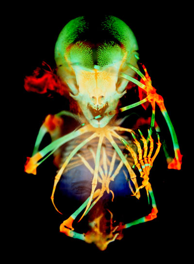 Fruit-bat embryo. Credit: Vanessa Chong-Morrison and Dorit Hockman
Course faculty Paul Trainor had brought a variety of embryos --- boas, bats, mice – for the students to stain for imaging however they wished. “I looked at Allan and we had the exact same thought – we had to try out the chameleon embryos, at least once in a lifetime,” says Salamanca. After bone staining the embryo, they imaged it on a microscope that Zeiss had loaned to the course.
“Actually, this picture was coincidental; not planned at all. We were checking the microscope’s channels, testing for the ideal color, and we found a fluorescent channel that perfectly showed the animal’s autofluorescence. You could see the surface of the skin together with the bones – simply beautiful,” Salamanca says. Since the sample was so big, they imaged several parts of the body and stitched them together afterwards. “Several photo contests were open at the time, and we thought, why not send it in,” he says. A favorable decision!
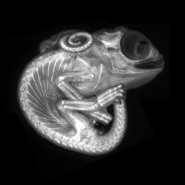 Chameleon embryo. Credit: Allan Carrillo-Baltodano and David Salamanca
Landing in 20th place in the Nikon contest is a fruit-bat embryo imaged by two MBL Embryology students who took the course three years apart. In 2015, students in the course were given a variety of preserved specimens so they could try out whole-mount skeletal stainings. Vanessa Chong-Morrison chose a fruit-bat specimen to prepare, and took the sample back with her to Oxford University, where she was a Ph.D. student. About a year later, she teamed up with a postdoctoral associate in the lab, Dorit Hockman (Embryology ‘12) to image the sample. Similar to the chameleon image, they took several shots of parts of the bat and stitched them together for the final result. “In hindsight, [waiting a year] contributed greatly to the high-quality resulting image, as skeletal stainings with regular washes increase in clarity over time,” Chong-Morrison says.
An exhibit of the winning images in the 2020 Nikon Small World Competition will be at the Marine Biological Laboratory from July 31 to October 1, 2021.