Every Photon is Valuable: New Microscope Captures “Lost” Fluorescence, Improving Resolution

Contact: Diana Kenney, dkenney@mbl.edu; 508-289-7139
WOODS HOLE, MASS.—Taking a cue from medical imaging, scientists have invented a multi-view microscope that captures higher-resolution, 3D images of live cells and tissues without upping the dose of potentially harmful radiation the specimens receive.
The researchers, who work collaboratively at the Marine Biological Laboratory’s Whitman Center, published their results this week in the journal Optica.
 Hari Shroff
“Everybody knows fluorescence imaging is inefficient in that the microscope only captures a portion of the light (spewing off the specimen),” says senior author Hari Shroff of the National Institute of Biomedical Imaging and Bioengineering. “In this paper, we showed you can not only capture that lost light, but use computation to fuse it to the existing image and make the image sharper.”
Developed by Yicong Wu, a staff scientist in Shroff’s lab, the new system achieved resolution of up to 235 x 235 x 340 nanometers, which is double the volumetric resolution of traditional fluorescence microscopy methods.
 Patrick La Rivière
To collect more of the available light (which, in turn, provides more information about the specimen), the new microscope has three objective lenses acquiring views of the sample simultaneously. The views are then aligned and merged by a computational process known as deconvolution.
Those computations were worked out in collaboration with co-author Patrick La Rivière of the University of Chicago’s Radiology Department, who typically develops algorithms for improving “dose efficiency” in human-scale medical imaging, such as CAT scans.
“In medical imaging, we are always worried about dose, about capturing every X-ray [used on the patient to improve scan resolution]. We are concerned with ‘How can we do more with less?’” La Rivière says.
In microscopy, the amount of light used presents similar concerns. “If you use very intense illuminations to image something microscopic like a worm embryo, you might change its biology or even kill it. You need to be dose efficient with your light,” La Rivière says.
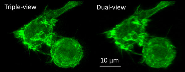 Macrophage actin labeled with green fluorescent protein, imaged with the new triple-view microscope (left) and the original dual-view microscope (the diSPIM, right). Credit: Yicong Wu and Valentin Jaumouille
La Rivière and Shroff first met at an MBL workshop on microscopy research in 2014. “On the plane [from Chicago], I read Hari’s paper about an earlier version of this microscope, and I saw he was using a very familiar, bread-and-butter deconvolution algorithm from the medical imaging world,” LaRivière says. “To me, that was the perfect entry point [into light imaging research]; it was something I knew. I chased Hari down after the workshop to see if he was open to collaboration, which he was.”
Supported by a University of Chicago-MBL Exploratory Research Fund award, the two began collaborating to improve Shroff’s microscope (the diSPIM, which has two objective lenses) and eventually the new three-lensed, triple-view microscope.
La Rivière this year was named an MBL Fellow. Shroff is a Whitman Center Scientist and co-director of the MBL’s Optical Microscopy and Imaging in the Biomedical Sciences course.
Citation:
Yicong Wu, P. Chandris, P.W. Winter, E.Y. Kim, V. Jaumouillé, A. Kumar, M. Guo, J.M. Leung, C. Smith, I. Rey-Suarez, H. Liu, C.M. Waterman, K.S. Ramamurthi, P. La Riviere, H. Shroff (2016) Simultaneous multi-view capture and fusion improves spatial resolution in wide-field and light-sheet microscopy. Optica 3, 8: 897-920; doi: 10.1364/OPTICA.3.000897
—###—
The Marine Biological Laboratory (MBL) is dedicated to scientific discovery – exploring fundamental biology, understanding marine biodiversity and the environment, and informing the human condition through research and education. Founded in Woods Hole, Massachusetts in 1888, the MBL is a private, nonprofit institution and an affiliate of the University of Chicago.