Images from the Embryology Course

This image of the arthropod Springtail (Collembola) was captured on a Flamingo T-SPIM lightsheet microscope. Credit: Guilherme (Gui) Gainett and Carsten Wolff.
Since 1893, students from around the world have congregated at the Marine Biological Laboratory for the popular Embryology Advanced Research Training Course. The students in this year's course, Embryology Concepts & Techniques in Modern Developmental Biology, have produced some eye-catching images during their time in Woods Hole.
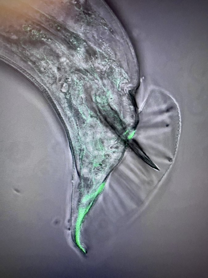
Male nematode (C. Elegans) spicules imaged on Zeiss Micro 900 with GFP laminin. Credit: Rebecca M Varney, Postdoc, UCSB.
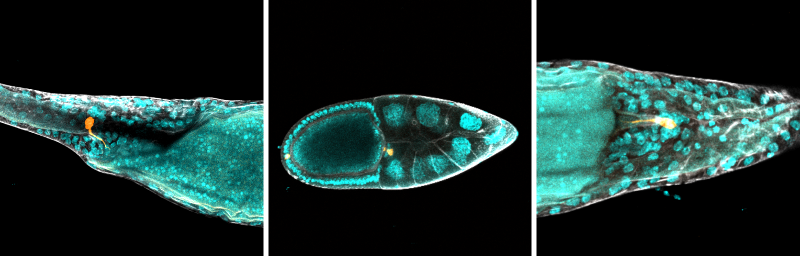
Egg chamber of fruit fly (Drosophila) imaged on a Zeiss LSM 900. Nuclei (cyan), F-actin (grey), polar cells (orange). Credit: Louis Prahl, postdoc, University of Pennsylvania
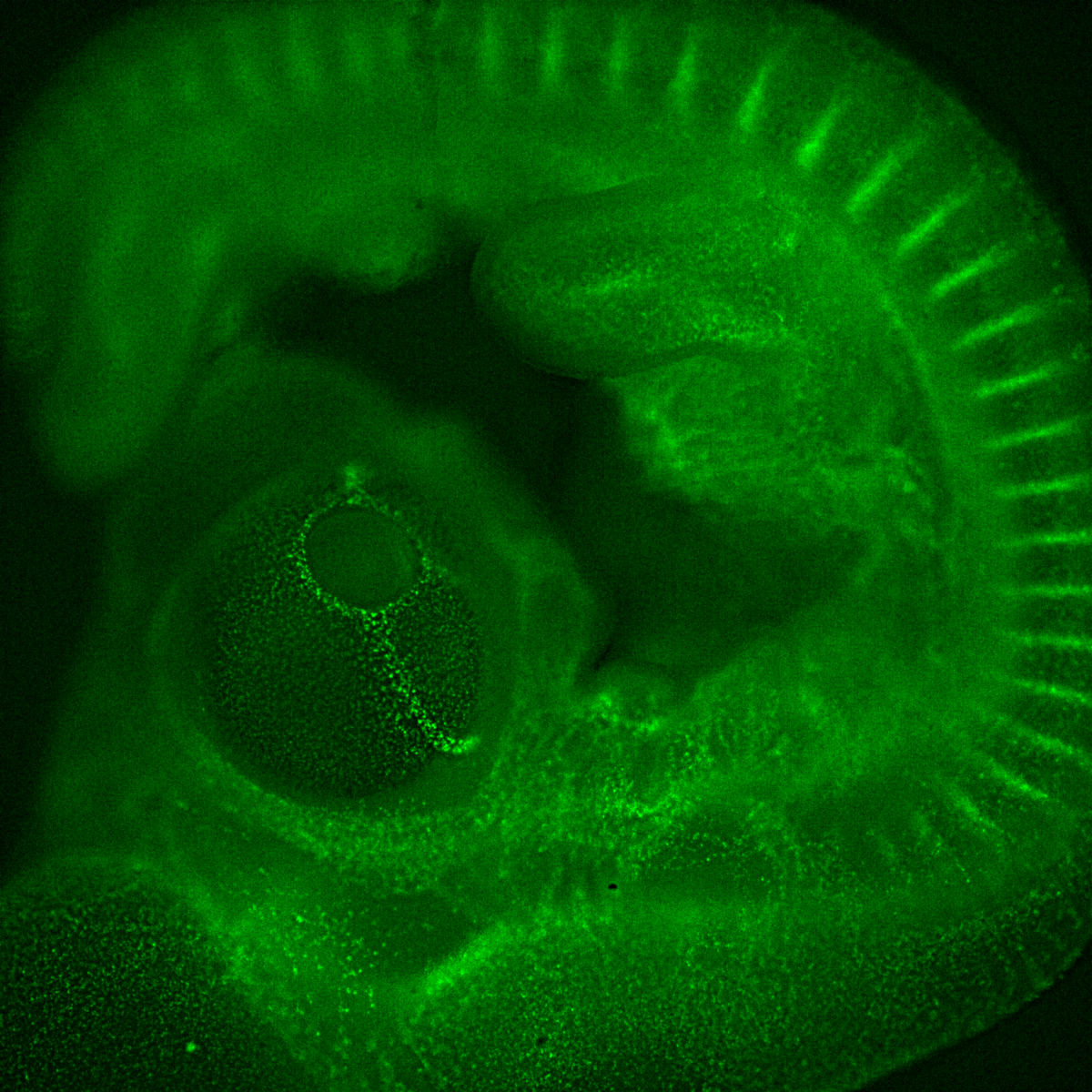
A transgenic quail from Peter Lwigale's lab, it is expressing fluorescence (GFP) to target vasculature, which you can see quite well around the eye at this stage. Imaged on a Leica Micro Thunder. Credit: Evan Curcio
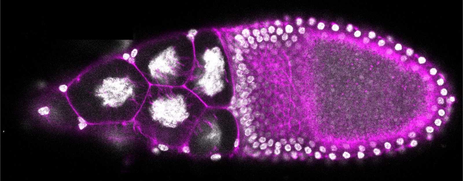 Fruit Fly (Drosophila) egg chamber. Actin filaments in magenta and nucleus/DNA in grey. Taken during #Embryo21 on ZEISS microscope. Credit: Viraj Doddihal, Graduate Student at the Stowers Institute for Medical Research, under direction of Sally Horne-Badovinac
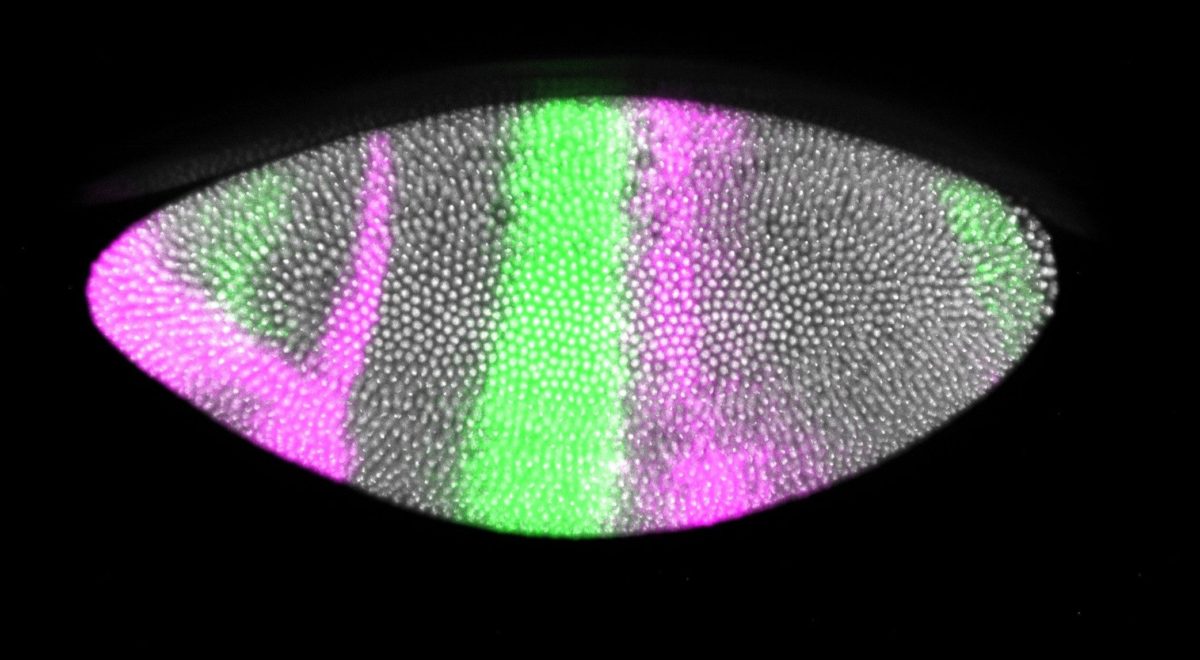 Early stage fruit fly (drosophila) embryos captured on a Nikon Ti2 microscope with a Yokogawa W1 spinning disk confocal. The colors are krüppel (green) and knirps (magenta) in situ hybridizations. Staining done by scientists in the Nipam Patel Lab. Credit: Louis Prahl, postdoc, University of Pennsylvania
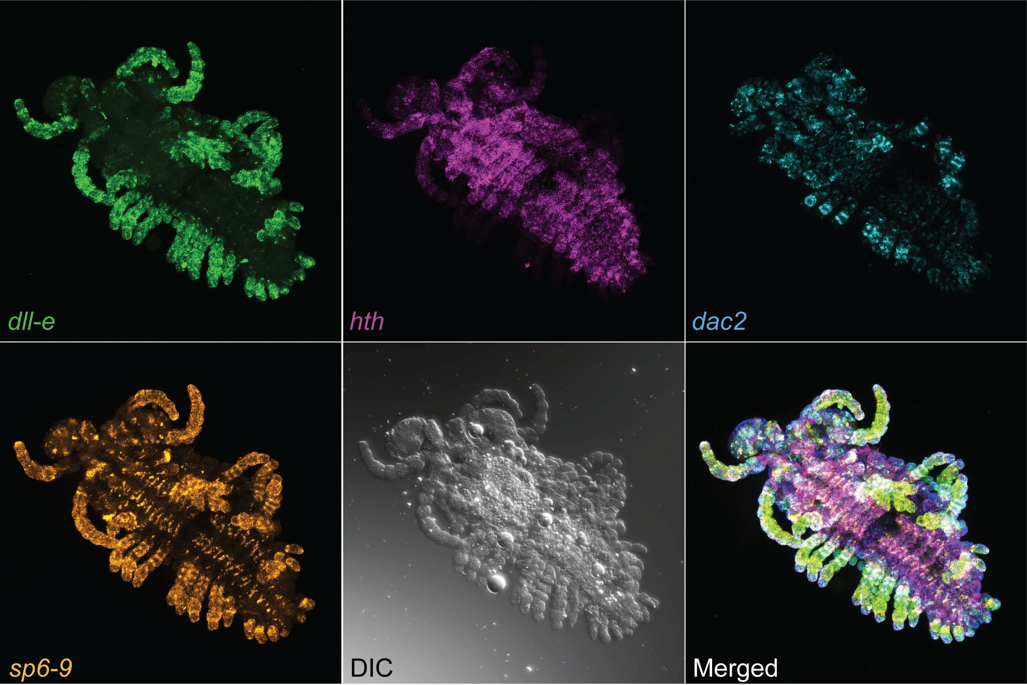 A set of lab gap genes in the amphipod crustacean (Parhyale hawaiensis). Genes expressed: dll-e (green), hth (purple), dac2 (blue), sp6-9 (orange), DIC (grayscale), Bottom right is a merged image showing all other at once. Credit: Brittany Truong, PhD Candidate, University of Colorado
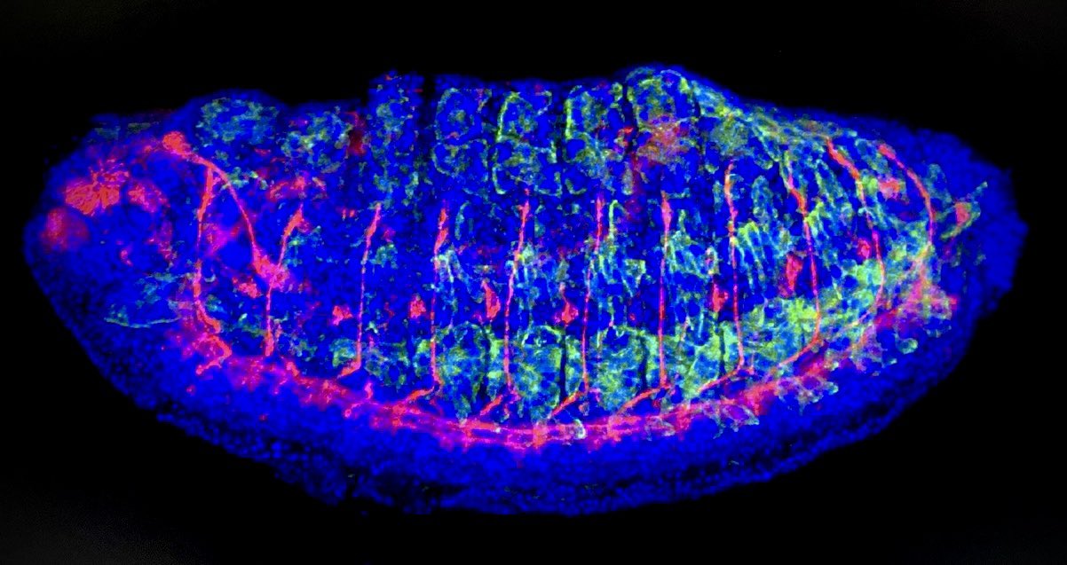 An embryo of the fruit fly (drosophila) imaged under a Zeiss 780 Confocal Microscope. Blue is DAPI for nuclei; red is motor neurons, green is muscle, and pink is central nervous system. Credit: Rebecca M Varney, Postdoc, UCSB.
For more microscopy images, follow #Embryo21 on Twitter.