Nobel Prize in Chemistry Awarded to Microscopist and MBL Visiting Scientist and Faculty Member Eric Betzig

FOR IMMEDIATE RELEASE
Contact: Diana Kenney, Marine Biological Laboratory
508-685-3525 (cell); 508-289-7139; dkenney@mbl.edu
WOODS HOLE, Mass.—When Eric Betzig loaded his hand-built, “bleeding edge” super-resolution microscope into an SUV in 2007 and drove it up to the Marine Biological Laboratory (MBL) for biologists to “test drive” it, it was immediately embraced as a major breakthrough in biological imaging. Today, Betzig received the Nobel Prize in Chemistry for development of that microscope technology, which allows one to discriminate individual molecules at nanometer resolution, far better than the previously presumed resolution limit of half the wavelength of light (0.2 micrometers).
Betzig is a group leader at Howard Hughes Medical Institute’s Janelia Research Campus and a recent MBL visiting scientist and faculty member. He has brought two new microscopes-under-development to the MBL, “where all sorts of world-class cell biologists throw everything they can think of at it. We can learn through trial by fire what works and what doesn’t,” Betzig says.
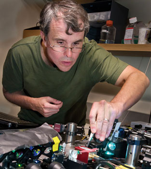 Eric Betzig tests one of his super-resolution microscopes, still under development, at the MBL. Credit: Tom Kleindinst
Also awarded the Nobel Prize in Chemistry today were Stefan W. Hell of the Max Planck Institute for Biophysical Chemistry and William E. Moerner of Stanford University. Due to the trio’s inventions, now known as nanoscopy, “scientists visualize the pathways of individual molecules inside living cells. They can see how molecules create synapses between nerve cells in the brain; they can track proteins involved in Parkinson’s, Alzheimer’s and Huntington’s diseases as they aggregate; they follow individual proteins in fertilized eggs as these divide into embryos,” the Nobel Foundation stated in its announcement today. “Theoretically, there is no longer any structure too small to be studied.”
Betzig and Moerner, working separately, laid the foundation for single-molecule microscopy. This method relies upon the possibility to turn the fluorescence of individual molecules on and off. Scientists image the same area multiple times, letting just a few interspersed molecules glow each time. Superimposing these images yields a dense super-image resolved at the nanolevel. Betzig used this method, called PALM (photoactivated localization microscopy), for the first time in 2006.
MBL Physiology course co-director Jennifer Lippincott-Schwartz of the National Institutes of Health made an important contribution to the development of PALM, and also brought Betzig to the MBL to test his new technology in the course. Writing about the collaboration in a 2008 issue of MBL Catalyst, Lippincott-Schwartz explained, “Typically, under a light microscope, fluorescently tagged molecules that are less than 200 nanometers apart blur into a fuzzy blob, so the individual molecules can’t be distinguished. Eric’s idea was to configure a light microscope so that only a small number of fluorescent molecules in the cell light up at once. After repeating this many times, the image layers could be combined into one, super-resolution image of the cell. To make this work, Eric needed a ‘photoconvertible’ fluorescent protein tag, meaning its illumination can be switched on or off at will.”
Just such a photoactivated tag had been created by George Patterson in Lippincott-Schwartz’s lab at the NIH, using green fluorescent protein (GFP). Betzig and Lippincott-Schwartz began collaborating in her lab, and within a few months the microscope prototype had been built, and they had proven the PALM technique worked.
“We were thrilled, but we didn’t stop there,” Lippincott-Schwartz wrote. “Initially, we were looking at fixed cells, but we knew scientists would really get excited if we could apply PALM to living cells. In the summer of 2007, I was invited to teach a two-week rotation in the MBL Physiology course. I invited Eric to accompany me, and he brought along Hari Shroff, who had been a Physiology student the year before. It was a fantastic experience. We worked around the clock, trying many different approaches. By the end of the rotation, we had gotten live-cell PALM to work! Not only that, we got PALM to work with two different colors of fluorescent probes, and we demonstrated that PALM could be used to track single molecules in live cells.”
Betzig has collaborated with MBL faculty and students in both the Physiology and Neurobiology courses, and served on the faculty of the Physiology course in 2011 and the Analytical and Quantitative Light Microscopy course in 2013.
Hari Shroff is also continuing the time-honed tradition of biologists and physicists interacting at the MBL to develop next-generation imaging instruments. Shroff, an investigator and microscope developer at the National Institutes of Health, co-directs the MBL’s Optical Microscopy and Imaging in the Biomedical Sciences course.
Betzig’s single-molecule technology was made possible by the discovery of green fluorescent protein (GFP) in the jellyfish Aequorea by MBL Distinguished Scientist Osamu Shimomura in 1961 at Princeton University. Shimomura was co-recipient of the 2008 Nobel Prize in Chemistry.
Resources:
To request an interview with Eric Betzig, please contact:
Jim Keeley, Howard Hughes Medical Institute
keeleyj@hhmi.org
301-215-8858
MBL Blog post (with videos) and HHMI Bulletin article about Betzig testing his new microscopes with MBL biologists.
Watch a video of Betzig giving the MBL Friday Evening Lecture, “Pushing the Envelope in Biological Fluorescence Imaging,” in 2012.
HHMI news release and background on Eric Betzig.
iBiology video, "Eric Betzig and Harald Hess: Developing PALM Microscopy"
Technical article introducing PALM technique: Betzig et al, Science 313: 1642-1645, 2006.
—###—
The Marine Biological Laboratory (MBL) is dedicated to scientific discovery and improving the human condition through research and education in biology, biomedicine, and environmental science. Founded in Woods Hole, Massachusetts, in 1888, the MBL is a private, nonprofit institution and an affiliate of the University of Chicago.