Scientists Propose Network of Imaging Centers to Drive Innovation in Biological Research

Contact: dkenney@mbl.edu; 508-289-7139
WOODS HOLE, Mass. -- When sparks fly to innovate new technologies for imaging life at the microscopic scale, often diverse researchers are nudging each other with a kind of collegial one-upmanship.
“Look at the resolution we obtain with this microscope I’ve designed,” the physicist says. “Great,” the biologist replies, “but my research organism moves fast. Can you boost the system’s speed?” “You’ll have terabytes of raw data coming off that microscope system,” says the computational scientist. “We’ll build in algorithms to manage the data and produce the most meaningful images.” Around they go, propelling a cycle of challenge and innovation that allows them to see more clearly into new dimensions of the microscopic world.
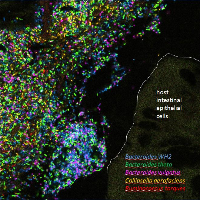 Bacteria in a model human gut microbiome established in a germ-free mouse. Credit: Jessica Mark Welch and Yuko Hasegawa
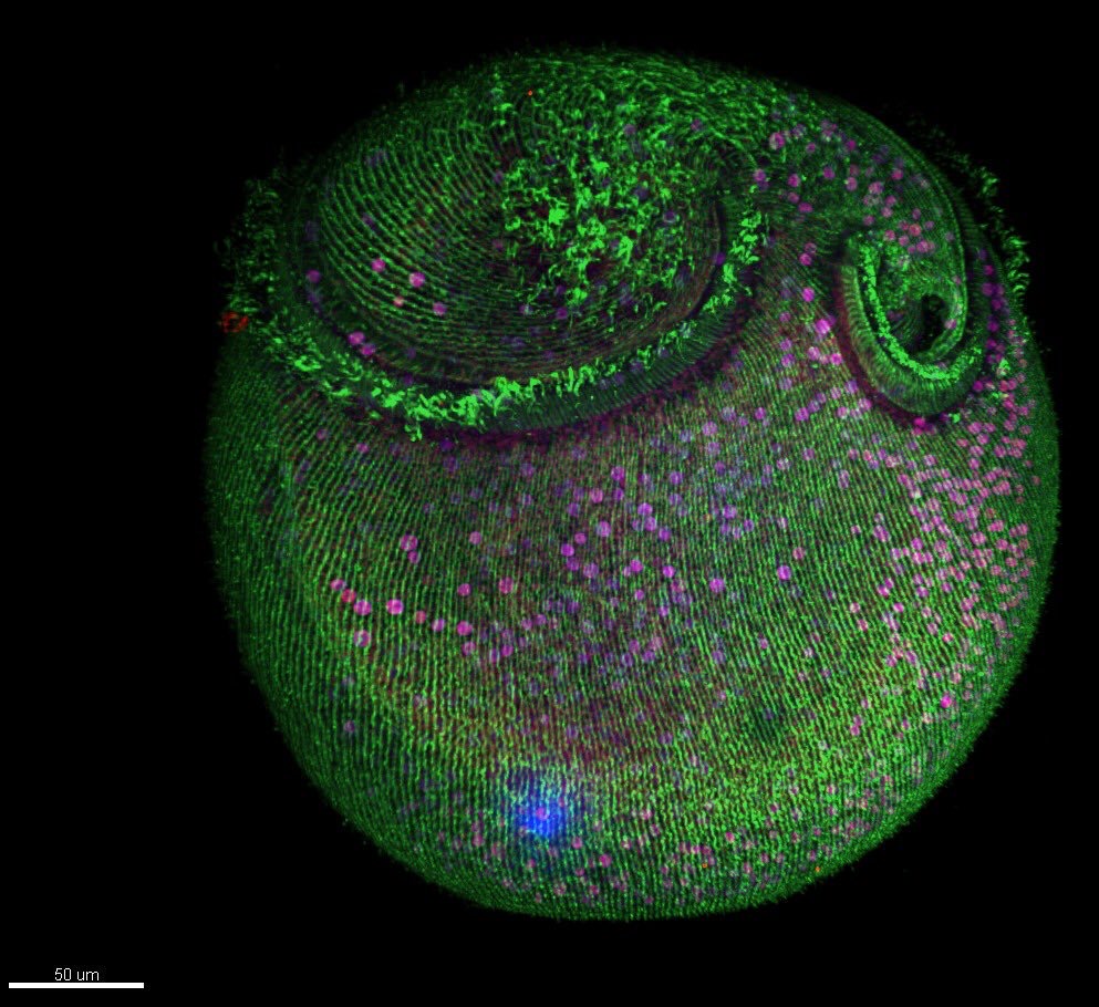 Microtubule structures in Stentor. Credit: Victoria Yan, MBL Physiology course
Interdisciplinary interactions are essential for driving the innovation pipeline in biological imaging, yet collegial workspaces where they can spring up and mature are lacking in the United States. Last fall, the Marine Biological Laboratory (MBL) convened a National Science Foundation workshop to identify the bottlenecks that stymie innovation in microscopy and imaging, and recommend approaches for transforming how imaging technologies are developed and deployed. The conclusions of the 79 workshop participants are summarized in a Commentary in the August issue of Nature Methods.
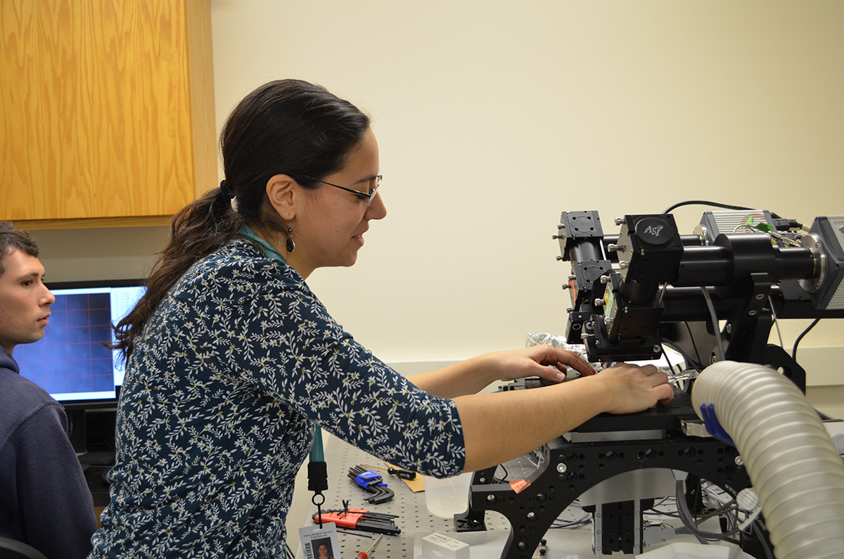 MBL Hibbitt Fellow Duygu Ozpolat learns how to use the diSPIM imaging system at an MBL workshop. Credit: Diana Kenney
“We propose a network of national imaging centers that provide collaborative, interdisciplinary spaces needed for the development, application, and teaching of advanced biological imaging techniques,” write the authors and workshop leaders, MBL Fellows Daniel A. Colón-Ramos of Yale University School of Medicine, Patrick La Riviere of the University of Chicago, Hari Shroff of the National Institute of Biomedical Imaging and Bioengineering, and MBL Senior Scientist Rudolf Oldenbourg.
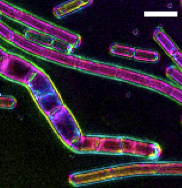 LC-PolScope image of cell wall sacculi purified from Bacillus subtilis with different levels of induction of mreBCD, the genes that build rod shape. Color indicates molecular orientation, brightness the degree of alignment. Credit: Dion, MF. et al (2019) Nature Microbiology.
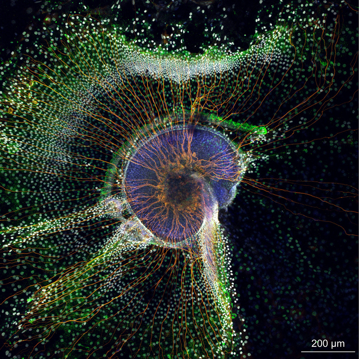 Mouse inner ear spiral ganglia culture. Credit: Mirko Scheibinger, MBL Biology of Inner Ear course
“Creating spaces for co-locating microscopists, computational scientists and biologists, in our experience, is a key and missing ingredient in new and necessary ecosystems of innovation. Sometimes the seeds of innovation have to be planted in the same pot for them to flower and be fruitful,” Colón-Ramos says.
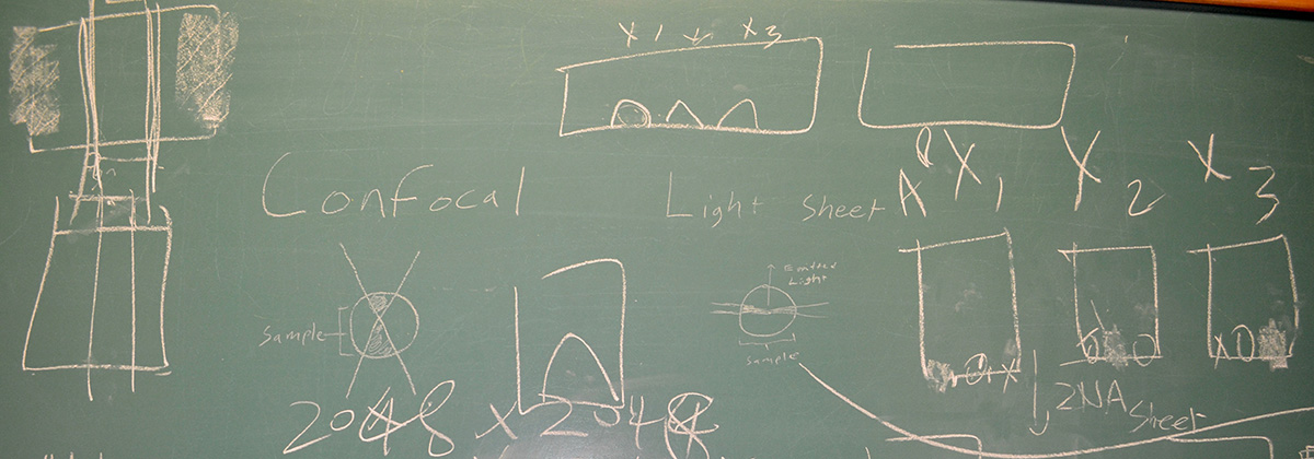 Blackboard notes comparing imaging systems in a MBL Whitman Center lab.
For the proposed centers to optimally succeed, they need expert staff scientists who engage in and catalyze collaborations among the major players in the imaging ecosystem, the group concluded. The centers will provide space and expertise not only for teaching existing, high-end imaging techniques, but also for designing and testing new technologies with real-world biological applications and disseminating them promptly to staff and visiting scientists, faculty and students. All can participate in an iterative innovation process, driving discovery while becoming interdisciplinary thinkers and project developers themselves.
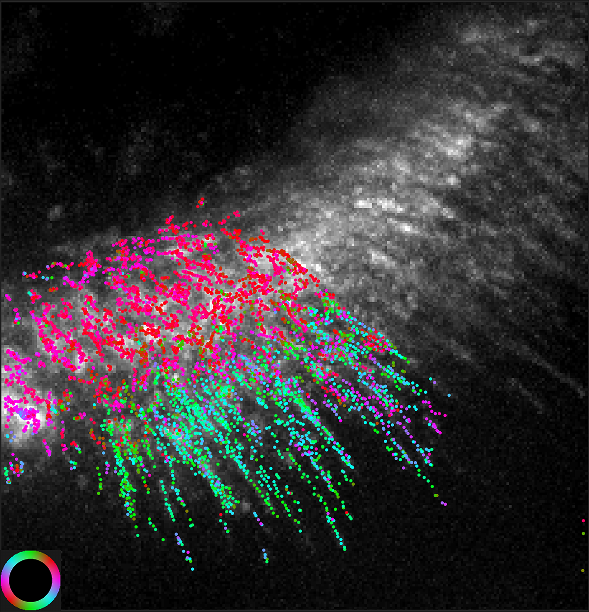 Color-coded actin filament orientation is overlaid on an image of fluorescence intensity. The orientation corresponds to the color wheel at bottom left. Credit: Shalin Mehta and Tomomi Tani
As a next step in transforming the vision of national imaging centers into a reality, a follow-up meeting with a variety of funding stakeholders will be scheduled in the spring of 2020. Ideas and helpful comments for the meeting may be sent to ImagingNetworks@MBL.edu.
An Imaging Initiative is in progress at the MBL, with many elements of the proposed national imaging centers already in place and others under development.
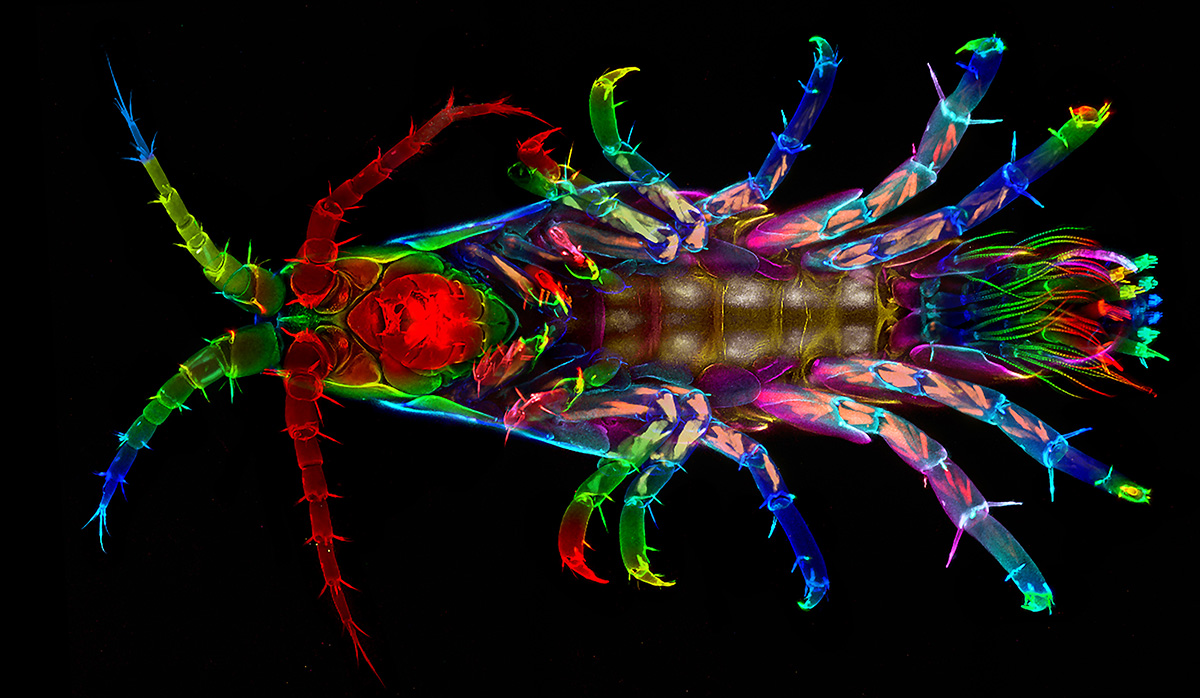 Parhyale hawaiensis, a model system for studying the evolution and development of body plans. Credit: Nipam Patel, MBL Embryology course
Citation: Daniel A. Colón-Ramos, Patrick La Riviere, Hari Shroff and Rudolf Oldenbourg (2019). Transforming the development and dissemination of cutting-edge microscopy and computation. Nature Methods, DOI: 10.1038/s41592-019-0475-y
Home page photo caption: Cuttlefish (Sepia officinalis) skin; neurons in red. Credit: Trevor Wardill
—###—
The Marine Biological Laboratory (MBL) is dedicated to scientific discovery – exploring fundamental biology, understanding marine biodiversity and the environment, and informing the human condition through research and education. Founded in Woods Hole, Massachusetts in 1888, the MBL is a private, nonprofit institution and an affiliate of the University of Chicago.