Vivid Microbial Landscape on the Human Tongue is Revealed

Contact: 508-685-3525; dkenney@mbl.edu
WOODS HOLE, Mass. – For the first time, researchers have developed high-resolution images of the microbial communities on the human tongue. The images, published this week in Cell Reports, reveal that microbial biofilms on the surface of the tongue have a complex, highly structured spatial organization.
“From detailed analysis of the structure, we can make inferences about the principles of [microbial] community growth and organization,” says senior author Gary Borisy, of the Forsyth Institute and the Harvard School of Dental Medicine. “Bacteria on the tongue are a lot more than just a random pile. They are more like an organ of our bodies.”
“We are looking at the mouth as a microbial landscape, where different bacterial communities occupy different kinds of habitats,” says co-author Jessica Mark Welch, a microbial ecologist at the Marine Biological Laboratory (MBL) in Woods Hole.
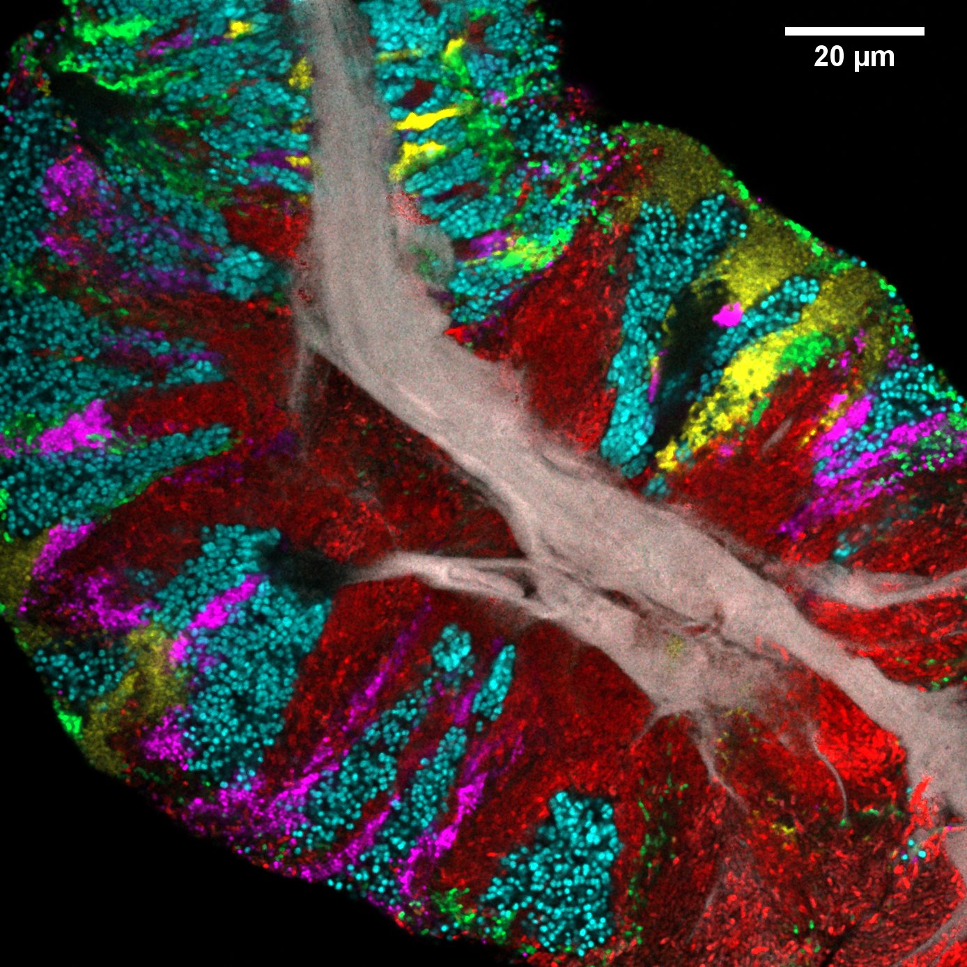 Bacterial biofilm scraped from the surface of the tongue and imaged using CLASI-FISH. Human epithelial tissue forms a central core (gray). Colors indicate different bacteria: Actinomyces (red) occupy a region close to the core; Streptococcus (green) is localized in an exterior crust and in stripes in the interior. Other taxa (Rothia, cyan; Neisseria, yellow; Veillonella, magenta) are present in clusters and stripes that suggest growth of the community outward from the central core. Credit: Steven Wilbert and Gary Borisy, The Forsyth Institute
“The tongue is particularly important because it harbors a large reservoir of microbes and is a traditional reference point in medicine. ‘Stick out your tongue’ is one of the first things a doctor says,” Mark Welch says.
The researchers used an imaging technique called Combinatorial Labeling and Spectral Imaging – Fluorescence in situ Hybridization (CLASI-FISH), which has been developed over the past decade in the Borisy lab and at the MBL. This strategy greatly expands the number of different kinds of microbes that can be simultaneously identified and localized in a single field of view.
“Our study is novel because no one before has been able to look at the biofilm on the tongue in a way that distinguishes all the different bacteria, so that we can see how they arrange themselves,” Borisy says.
“We think that learning who is next to who will help us understand how these communities work,” Mark Welch says.
The researchers identified 17 bacterial genera that were abundant on the tongue and present in more than 80 percent of the samples (taken from 21 healthy individuals). The samples all contained bacteria organized into consortia (structurally complex, multi-layer biofilms) as well as free bacteria and bacteria bound to host epithelial cells.
“What was striking is we observed the same microbial [consortia] on the tongue of every person we sampled,” Mark Welch says. These consortia contain abundant cells of three genera – Actinomyces, Rothia, and Streptococcus – arranged structurally in similar ways.
Their results suggested a model for how microbial community structure on our tongues is generated. First, bacterial cells attach to the epithelium of the tongue’s surface singly or in small clusters. During population growth, differing taxa push on one another and proliferate more rapidly in microenvironments that support their physiological needs. This differential growth results in the patch mosaic organization observed in larger, more mature structures on the tongue.
While the function of the oral microbiome is not yet understood, the images also revealed the presence of several genera capable of nitrate reduction in saliva, a metabolic transformation that is likely important to human health but is not encoded by the human genome.
Mark Welch’s lab has extended the CLASI-FISH technology to image a variety of microbial communities: in colon cancer patients; in marine organisms including the cuttlefish; and on microplastic ocean debris. She and Borisy have previously collaborated to image the microbial structures in dental plaque and in a model human gut microbiome.
This work was supported by the National Institutes of Health.
Citation: Steven A. Wilbert, Jessica L. Mark Welch and Gary G. Borisy (2020) Spatial Ecology of the Human Tongue Dorsum Microbiome. Cell Reports, DOI: 10.1016/j.celrep.2020.02.097
—###—
The Marine Biological Laboratory (MBL) is dedicated to scientific discovery – exploring fundamental biology, understanding marine biodiversity and the environment, and informing the human condition through research and education. Founded in Woods Hole, Massachusetts in 1888, the MBL is a private, nonprofit institution and an affiliate of the University of Chicago.