Creature Feature: Research Organisms of the MBL

By Emily Greenhalgh
The organisms studied by MBL scientists may not be spooky or scary, but some of them are downright weird. That’s what makes them such great research subjects.
Below are just a few of the amazing organisms scientists are working with at the MBL.
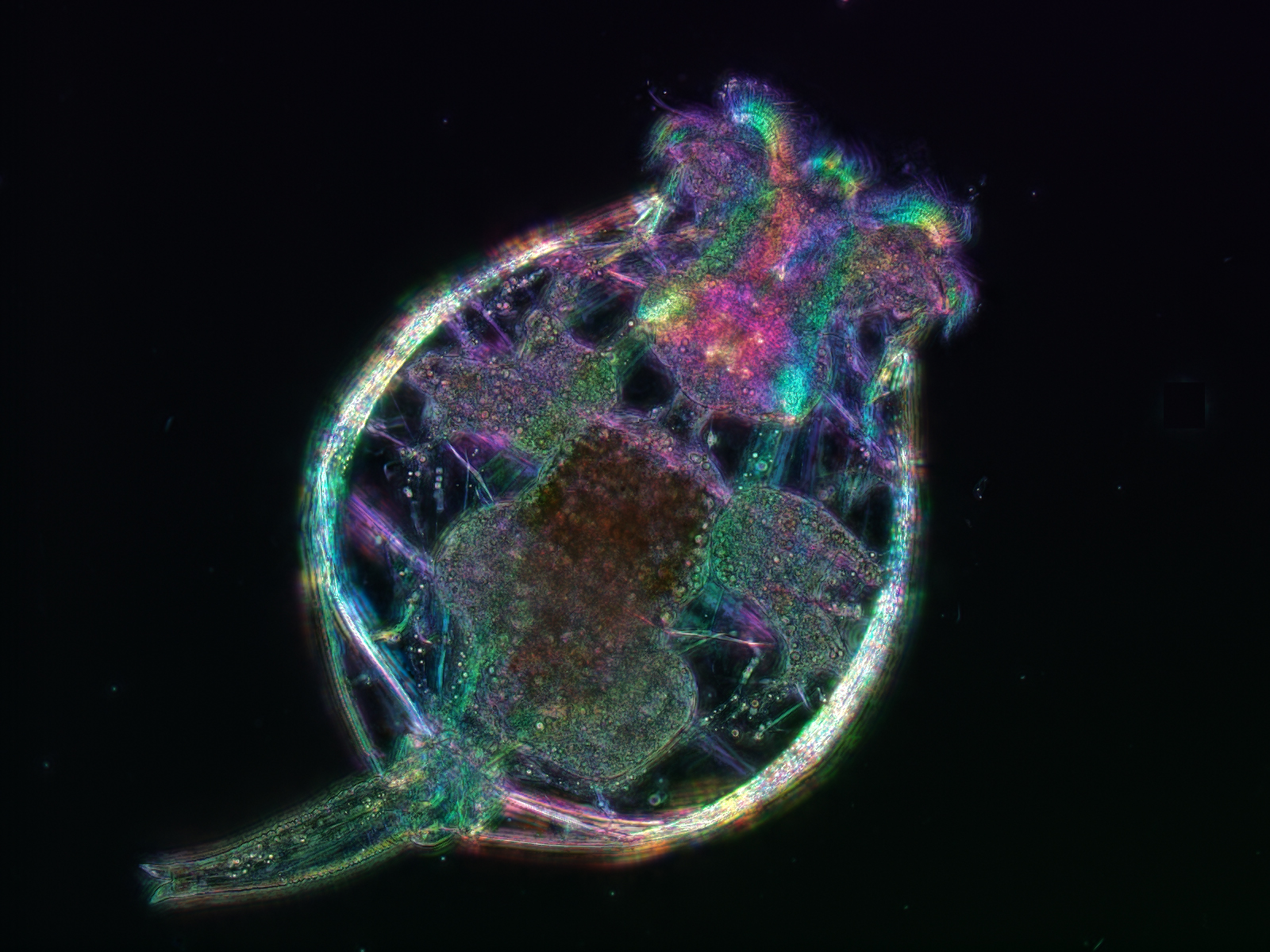 Rotifer. Credit: Kristin Gribble and Michael Shribak | Equipment: Polychromatic Polscope
Rotifer: Small, but Mighty
For scientists at the MBL, the rotifer (Brachionus manjavacas) is used as a model organism to study evolution, stress responses, the biology of aging, and maternal effects. Rotifers are small, easy to grow in the lab, have a short lifespan, and share many of their genes with humans. That makes them ideal specimens in which to address questions relevant to human health as well as understand basic biological and evolutionary processes. Brachionus rotifers produces eggs that can be completely dried and frozen for decades, then hatch within a day when exposed to water and light.
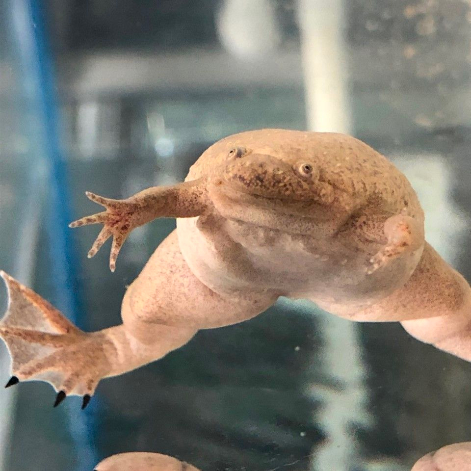 Xenopus tropicalis. Credit: James Parente | Equipment: iPhone 8
Xenopus: Mutant Madness
At the National Xenopus Resource (NXR) at the MBL, there are thousands of western clawed frogs (Xenopus tropicalis), including many mutant and transgenic lines. It’s the only species in the Xenopus genus to have a diploid genome, making it ideal for genome editing. They’re widely used in research thanks to a powerful combination of experimental tractability and a close evolutionary relationship with humans. These frogs are used by MBL’s year-round and visiting researchers for a wide variety of research topics including sex determination, modeling genetic diseases, as well as developmental and evolutionary biology.
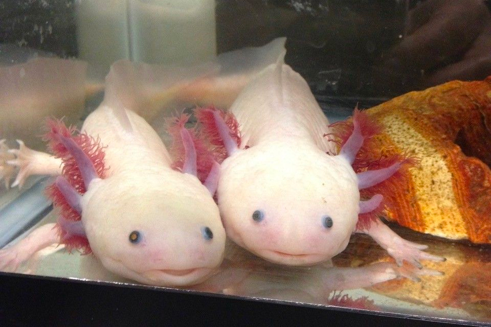 Two axolotls in a tank. Credit: Karen Echeverri | Equipment: iPhone
Axolotl: Superhero of Regeneration
The Axolotl (Ambystoma mexicanum) can functionally regenerate multiple body parts without forming scar tissue. MBL scientists are studying these animals hope to decipher how the cells in these animals respond to injury in the spinal cord, limbs, and skin at the cellular and molecular level, and how that process differs from healing in humans, who can’t regenerate. Researchers in the MBL’s Echeverri Lab are identifying critical molecules, regulatory pathways and cellular processes in the axolotls underlying scar-free regeneration.
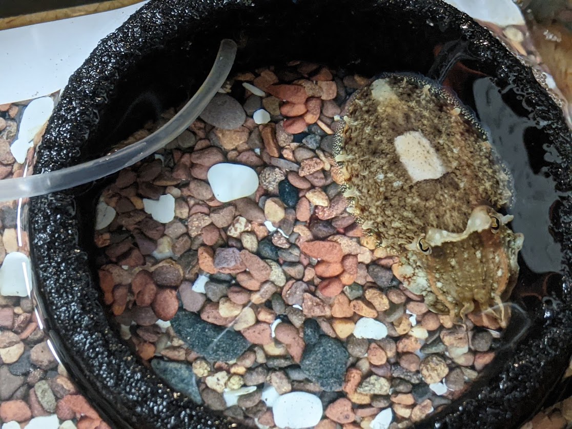 Common cuttlefish showing patterning to match rocks in Roger Hanlon Lab at MBL. Credit: Emily Greenhalgh
Common Cuttlefish: Masters of Camouflage
Their name may be common, but the common cuttlefish (Sepia officinalis) is a master of camouflage. Part of the cephalopod family that includes squid, octopuses, and nautiluses, cuttlefish’s camouflage abilities are due to special color-changing organs in their skin called chromatophores. By changing the patterns and backgrounds around the cuttlefish, scientists in the Roger Hanlon Lab, are investigating how well S. officinalis can change the color, pattern, contrast, and 3D physical texture of their skin to match their surroundings.
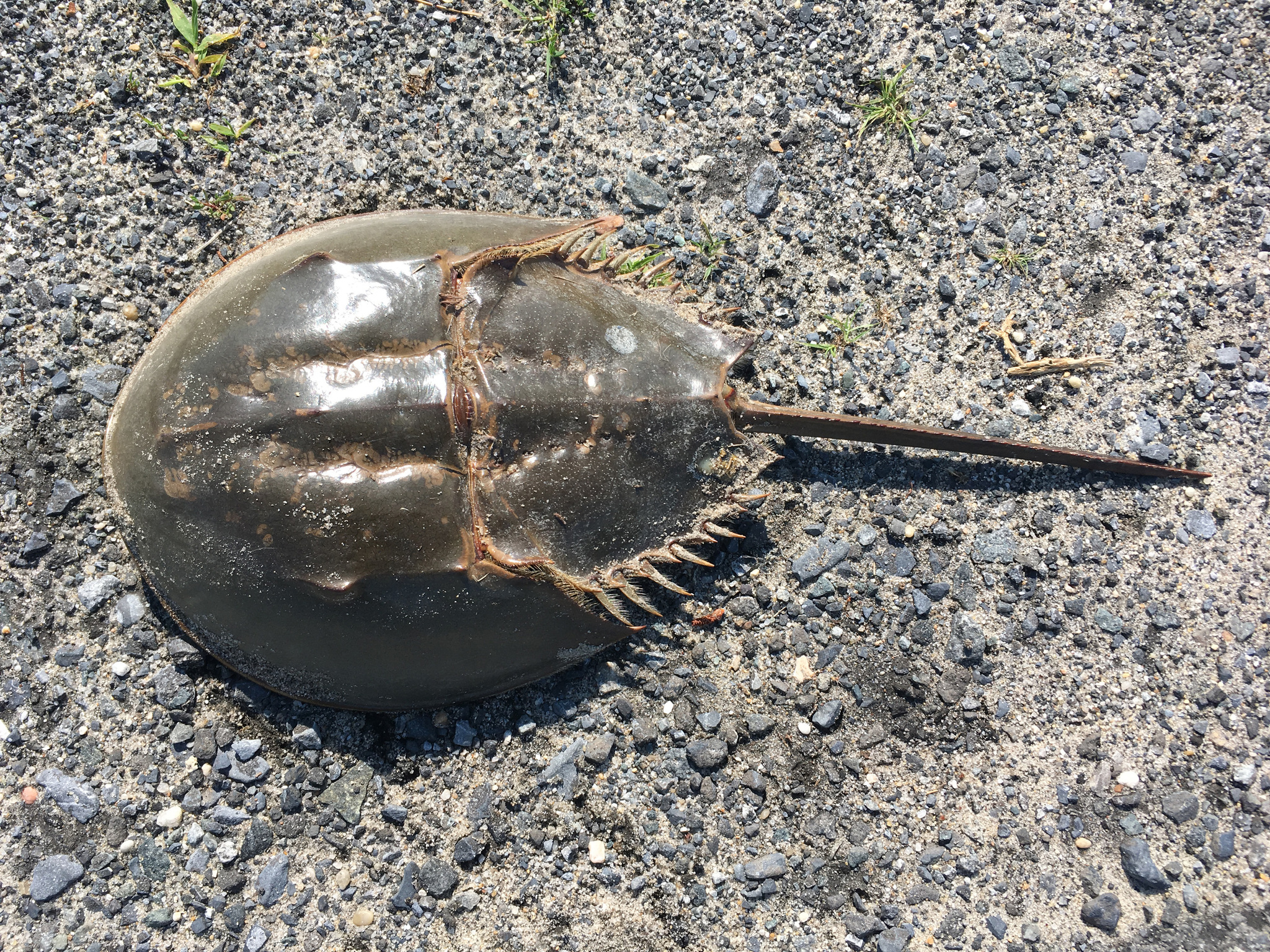 Atlantic horseshoe crab (Limulus polyphemus) on beach. Credit: Wikimedia Commons
Horseshoe Crab: Living Fossil
The horseshoe crab (LImulus polyphemus) has been on Earth more than 445 million years—longer than the dinosaurs. The organisms that survive today are almost identical to their ancient ancestors. The characteristic blue blood of horseshoe crabs is used in biomedical research to test pharmaceuticals and medical devices for potentially deadly endotoxins. This test, called the Limulus Amebocyte Lysate (LAL) takes advantage of the horseshoe crab’s primitive immune system, which causes its blood to clot when it encounters potentially harmful gram-negative bacteria. These living fossils are prized by MBL researchers studying immunity and also vision.