Gentle Mounting Gel Assists in Imaging of Wriggly, Live Organisms, Team Reports


It was a classic MBL encounter: Two scientists with nothing yet in common bump into each other on the campus, sparks fly, and within minutes they’ve dreamed up a new collaboration.
During a brief, inspired conversation last summer, MBL biologist Eric Edsinger and bioengineer Dirk Albrecht of Worcester Polytechnic Institute realized they had a “win-win” experiment to try – one that would be “fun and easy,” too. The outcome of that fruitful collaboration was published last month in Nature Communications Biology.
The paper addresses a conundrum in microscopy: When imaging a live organism, how do you keep it from wriggling all over or escaping the field of view, without perturbing the very biological process you want to study?
Albrecht had discovered that an absorbent, optically clear gel called PEG hydrogel could offer a great solution for gently immobilizing worms (C. elegans) in a hydrated and controllable environment, allowing continuous imaging for hours without damaging the worms.
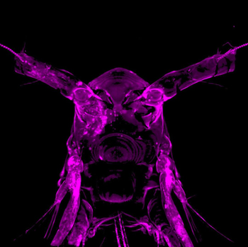 Live plankton (copepod) in PEG hydrogel, imaged with confocal microscopy. Credit: Kirsten Peramba, Peter Newstein and Eric Edsinger
Albrecht brought his gel to the MBL last summer to share lab space with MBL Fellows Hari Shroff and Daniel Colón-Ramos, who are engaged in a long-term collaboration to image the developing nervous system of C. elegans. Very soon, word of the wonderful gel spread. Scientists started bringing over their samples – jellyfish, hydra – to encapsulate in the gel and mount on a light-sheet microscope invented by Shroff.
Edsinger heard about the gel “through the grapevine” and walked over to Albrecht’s lab to check it out. He was impressed. “This gel is so simple. You just make a solution, put in your organism, hit it with UV light for a few seconds, and the gel forms around your sample. We might be able to take just about anything out of the ocean and mount it on many different microscopes using this gel! It’s really exciting, in that sense.”
Albrecht told him he was preparing a manuscript on the gel’s effectiveness for imaging wriggly C. elegans worms. “Maybe we can try scaling it to other organisms,” he said to Edsinger. “It sounds like different organisms have different mounting and imaging needs.”
“Yes, different bottlenecks,” Edsinger said. “What we can do is test the gel on various plankton that have different modes of locomotion. Crustaceans, gastropods, different ciliated types, jet propulsion types. Plankton is fun, anyways. And we can try it with my organism, pygmy squid."
“Alright, let’s do it! ” Albrecht said. So for the next few days they tested the gel with a variety of organisms. The final paper reports on the gel’s utility with C. elegans and pygmy squid, but the gel was tested on many other systems, from fruit flies to zebrafish, demonstrating its scaleability to sample sizes from larva to adult and its easy compatibility with a variety of microscope mounting geometries.
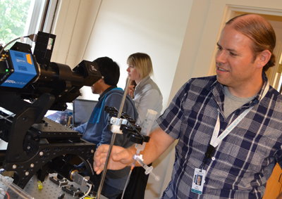 Dirk Albrecht at MBL working with the diSPIM microscope. Credit: D. Kenney
Among the many advantages of the PEG hydrogel is that its refraction index is similar to water. “That’s very important for imaging,” Colón-Ramos says. “It’s basically like the organism is surrounded by water, but the animal can’t move and the optical properties are very good,” Albrecht says.
Seawater native to the organism, nutrients or chemicals can be added to the porous gel and they diffuse quickly through it. The gel’s stiffness, and therefore how much it constrains movement, can be controlled by the concentration of the polymer. Organisms appeared healthy inside the gel: Squid hearts beat and their skin chromatophores open and close. Even after 24 hours in the gel, the paper reports, most of the young adult C. elegans crawl away when they are released.
Edsinger is now testing the gel with squid embryos to see if it can be used for developmental time-lapse imaging. “Development is sensitive to physical forces, and embryos want to change their shape as they go through morphogenesis. So we don’t know if this gel will change that biology,” he says. “But the great thing about it is you have a lot of control over how soft or rigid the gel is. We are just starting to explore tuning the gel to different phases of embryonic development. That’s this summer’s work!”
Citation: Kyra Burnett, Eric Edsinger and Dirk Albrecht (2018) Rapid and gentle hydrogel encapsulation of living organisms enables long-term microscopy over multiple hours. Communications Biology, DOI: 10.1038/s42003-018-0079-6