Legends of Squid Research at the MBL: Chasing Down Why Neurons Live or Die

Fresh ideas propel scientific discovery, but so do inventions, as the career of Elizabeth ‘Liz’ Jonas vividly shows. And Jonas’s invention happened in classic Marine Biological Laboratory (MBL) style: with salvaged sculpture wire, purple tape, and dashes of ingenuity, collaboration and laughter. It enabled years of future research that eventually led her to close in on an elusive accomplice in cell death associated with many serious disorders, including neurodegenerative disease.
Jonas was an undergraduate at Yale in 1980 when she got the chance to spend a summer at the MBL observing neuroscientist Rodolfo Llinas of New York University. Llinas was describing the properties of neurotransmission — the electro-chemical conversation that passes between neurons — using Woods Hole squid as his research organism. In squid, there is one communication junction between neurons that is “giant”– it’s 200 times larger than the largest human synapse — making this an ideal organism for studying neurotransmission. (See animation.)
“It was a really exciting summer,” Jonas says. “The squid boat (then the Loligo; today it’s the Gemma) would roll in between 11:30 AM and 3 PM with the day’s catch, and then the scientists would stay up all night doing experiments. I learned a tremendous amount and I just loved every minute of it.”
Jonas was hooked. After earning her M.D. and completing her residency training in Neurology, Jonas joined the lab of Leonard (Len) Kaczmarek at Yale as a postdoctoral scientist. Here, she worked with Kaczmarek on cutting-edge investigations of the class of proteins called ion channels that regulate a neuron’s excitability, or its ability to conduct charge. Kaczmarek co-directed the MBL Neurobiology course for five summers in the early ‘90s.
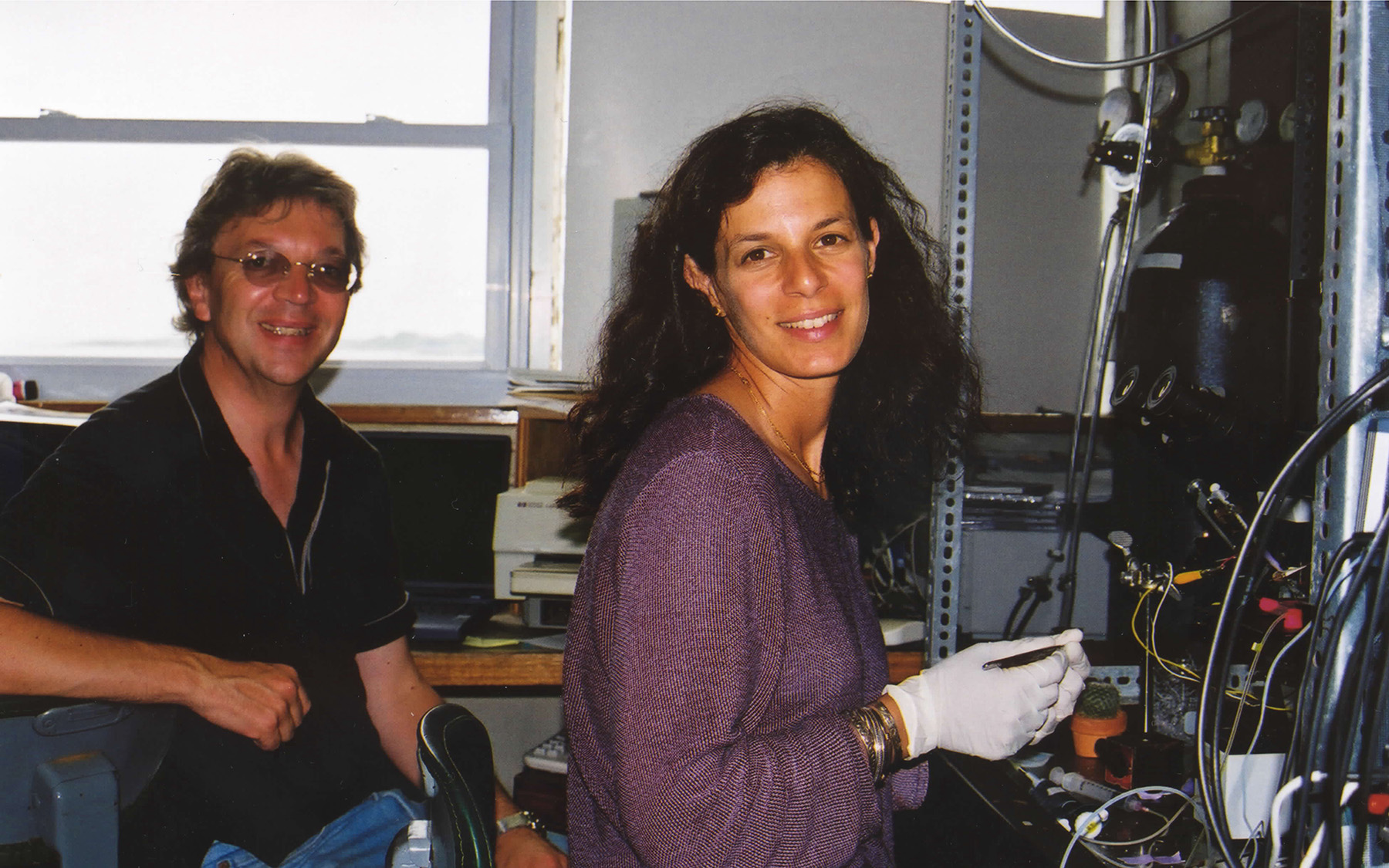 Len Kaczmarek and Liz Jonas of Yale University in their Whitman Center lab at MBL in 2001. Credit: J. Marie Hardwick
A Crazy Idea
The next summer, Jonas had what she calls a “crazy idea” and convinced Kaczmarek to return to the MBL Whitman Center to explore it. “It wasn’t difficult,” says Kaczmarek. “My family wanted to come back. My kids had grown up here. They love Woods Hole.”
Jonas’s idea? Rather than recording electrical current across the neuron’s outer membrane, Jonas wanted to try and record conductances inside the neuron — from the membranes of its tiny, internal organelles.
“We started working on this at Yale, but we realized that a nice preparation in which to try this would be squid,” Jonas says. And the only place to get squid was the MBL.
Once back in Woods Hole in an MBL lab, they zoomed in on recording from organelles inside the squid presynaptic terminal, where neurotransmitter is packaged for release.
Jonas had a problem, however. Her idea called for two electrodes that move relative to each other, but she couldn’t find a way to apply the force needed for them to move.
“We finally solved that problem using a piece of sculpture wire that Len’s wife had as part of her art supplies,” Jonas said, “and some purple tape. Steven Zottoli, a neuroscientist at MBL from Williams College, subsequently claimed that it was his roll of purple tape! He put up notices all over the lab stating he had invented the Purple Tape Wire. That became a playful bone of contention — Now we’d have to get the legal teams involved! We had a lot of fun with that.”


The Life and Death of a Neuron
Jonas’s technique opened a whole new world of inquiry inside neurons. She began recording from “very unusual, large-conductance ion channels” within the squid presynaptic terminal. With Joanne Buchanan at MBL, she eventually found that the recordings were coming from mitochondrial membranes. In nerve cells, mitochondria supply the energy for neurotransmission, among other functions. Most intriguingly, it was known that if a particular channel on the mitochondrial membrane remains open too long — in any type of cell — the cell will die.
“Various kinds of stresses, such as the lack of blood supply known as ischemia, can open this channel,” says Jonas. “It’s involved in diseases of the nervous system, heart, kidney, liver, muscle, pancreas; in diabetes, cancer – almost any organ you can think of,” she says.
But at the time nobody knew – and it’s still debated today – what membrane proteins form this important channel, known as the mitochondrial permeability transition pore (mPTP).
“It’s one of the Holy Grails of mitochondrial biology. What is the molecular identity of the mPTP?” says J. Marie Hardwick of Johns Hopkins Medicine, whose lab studies cell suicide and its role in disease.
Cell suicide (programmed cell death) is part of the normal development of an organism, as unneeded cells are pruned out. But in neurodegenerative disease, “cells die when they aren’t supposed to. The other side of that is cancer, when tumor cells manage to avoid cell death,” Hardwick says.
Jonas joined the Yale faculty in 2002 and dedicated a portion of her lab work to tracking down the ion channel associated with the mPTP. Soon after, Hardwick began coming regularly to MBL to collaborate in the quest, at the suggestion of her and Kaczmarek’s colleague, John Hickman. Hardwick is particularly interested in a family of proteins, called Bcl-2, known to regulate cell death.
“It took three summers before we could publish our first joint paper on this, because the squid weren’t available anywhere else,” says Hardwick. “We had a big tub of squid in the lab and many messy experiments.” In true MBL fashion, they collaborated with each other and MBL technical staff to supply materials and equipment, perform experiments and interpret the data. “Those were very fun times,” Hardwick remembers.

The team identified a candidate for the mPTP in 2011 and 2014, though their findings were controversial.
“Liz came under fire for this, but my perception now is that a lot of people are paying attention to it. Liz gets it right. She is a very daring scientist,” Hardwick says.
“We know for a fact that we’ve identified the largest-conductance channel on the mitochondrial inner membrane to date. But at this point, to say this is the only mPTP would be incorrect,” says Jonas. “There are many channels, and they are all participating in permeability of the membrane. The question is: Which are involved in the complex phenomenon of mitochondrial permeability transition, and how do those channels work together?”
Jonas’s lab is actively collaborating to develop drugs to close the mPTP under pathological conditions, as are other labs around the world.
Equally important, Jonas’s lab has unearthed surprising fundamentals about the role of the mPTP.
“Not only is it a cause of cell death, we’ve discovered it’s a major regulator of metabolism,” she says. “It regulates body weight, temperature, and the metabolic switches that can activate cells.” Jonas published her first big paper on regulation of neuronal developmental metabolism by the putative mPTP in Cell, 2020.
MBL as a Second Home
While Jonas’s lab stopped using squid several years go (it now uses mammalian systems), she and Kaczmarek still spend every summer at MBL “because we love it here,” Jonas says. They and their families have raised children here, and they’ve trained countless students in their MBL lab. Many of their students learn electrophysiology (a technique few labs now perform), but also cell imaging. These students are learning “from the best,” Hardwick says.
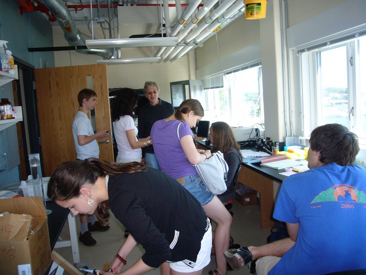 The Jonas-Kaczmarek-Hardwick lab at MBL is often bursting with student trainees, as shown in this photo from 2006. “We’ve had as many as 10 or 11 students in our lab” over the course of a summer, says Jonas. Credit: J. Marie Hardwick
 Liz Jonas, Steve Zottoli (center, trustee of the Grass Foundation/Grass Lab at MBL), and Len Kaczmarek with students in 2006. Credit: J. Marie Hardwick
“What’s exciting about electrophysiology is you see the brain working,” says Kaczmarek. “You see channels opening, neurons firing, ensembles of neurons. And you see your results during the experiment.”
Their labs enjoy taking multiple approaches: electrophysiology, imaging, biochemistry, genetics and computation. “We believe that if you want to solve a problem, you’ve got to find the technique that will help you solve it,” Jonas says.
Hardwick also comes back to MBL each summer, family in tow, to continue her fruitful collaborations. In recent years, she has taken a keen interest in whether single-celled organisms, such as yeast and microbes, also undergo cell suicide – a provocative idea that has caught the interest of other scientists at MBL.
“The MBL enables you to really appreciate collaborations, teaching and mentoring,” Jonas says.
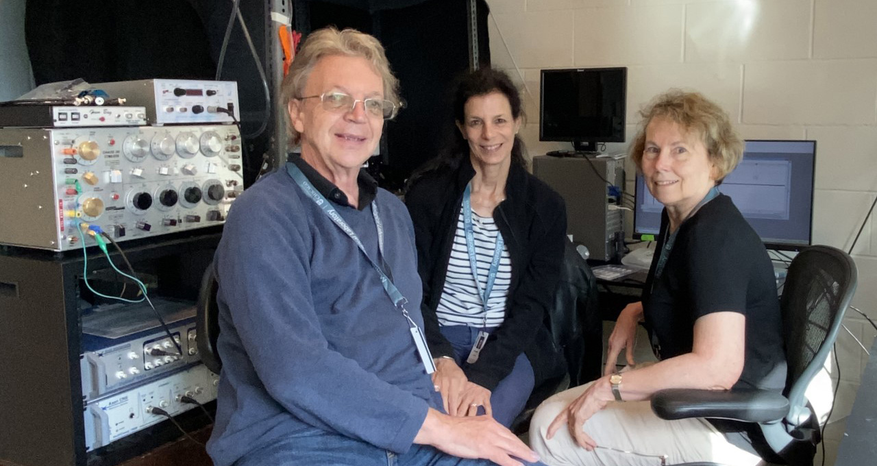 Len Kaczmarek and Liz Jonas of Yale University and J. Marie Hardwick of Johns Hopkins Medicine in their Whitman Center lab at MBL in 2021. Photo courtesy of J. Marie Hardwick
References:
Jonas EA, Buchanan J, Kaczmarek LK (1999) Prolonged activation of mitochondrial conductances during synaptic transmission. Science. doi: 10.1126/science.286.5443.1347.
Jonas EA, Hickman JA, Hardwick JM, and Kaczmarek LK (2004) Exposure to hypoxia rapidly induces mitochondrial channel activity within a living synapse. J. Biol. Chem. doi: 10.1074/jbc.M410661200
Jonas EA et al (2004) Proapoptotic N-truncated BCL-xL protein activates endogenous mitochondrial channels in living synaptic terminals. Proc. Natl. Acad. Sci. doi: 10.1073/pnas.0401372101
Chen YB et al (2011) Bcl-xL regulates mitochondrial energetics by stabilizing the inner membrane potential. J. Cell Biol. doi: 10.1083/jcb.201108059
Hardwick JM, Chen Y and Jonas AE (2012) Multipolar functions of BCL-2 proteins link energetics to apoptosis. Trends Cell Biol. doi: 10.1016/j.tcb.2012.03.005
Alavian KN, et al (2014) An uncoupling channel within the c-subunit ring of the F1FO ATP synthase is the mitochondrial permeability transition pore. Proc Natl Acad Sci U S A doi: 10.1073/pnas.1401591111
Kaczmarek LK, Zhang Y (2017) Kv3 channels: Enablers of rapid firing, neurotransmitter release, and neuronal endurance. Physiol Rev. doi: 10.1152/physrev.00002.2017.
Kulkarni M, Stolp ZD and Hardwick JM (2019) Targeting intrinsic cell death pathways to control fungal pathogens. Biochem. Pharmacol. doi: 10.1016/j.bcp.2019.01.012
Licznerski P et al (2020) ATP Synthase c-subunit leak causes aberrant cellular metabolism in Fragile X Syndrome. Cell, doi: 10.1016/j.cell.2020.07.008