Maienschein Lab Receives NSF Award to Study History of Cell Biology and Images

In 2018, the National Science Foundation (NSF) program “Understanding the Rules of Life: Building a Synthetic Cell” held an Ideas Lab to generate projects to develop synthetic cells. MBL Fellow Jane Maienschein attended. She became part of a team whose goal is to develop artificial cytoskeletons to place in living cells. In other words, the team will create a kind of “cyborg cell.” MBL Whitman Center scientist Fred Chang and University of Chicago modeler Aaron Dinner are co-principal investigators on this project, along with Mary Elting at North Carolina State (PI) and Saad Nahmla at Georgia Tech.
The project members will tackle central underlying questions about rules of life: What is a cell? How do we know when a synthetic unit counts as a cell? What is the history of cell biology that has led us to the assumptions about cells we hold today?
Maienschein, Karl Matlin (MBL Whitman Center/University of Chicago) and Manfred Laubichler of Arizona State University and Santa Fe Institute then received an additional NSF grant at the MBL to address those questions, Besides conducting historical investigations, Matlin and Maienschein will hold a set of workshops at the MBL. Those workshops will focus especially on how scientists created images of cells in the past, and how they used those images to present their ideas about cells through time.
In 2021, an artist and graduate student studying the history of biofilm research will be in residence at the MBL to help with the project. Working closely with Matlin and MBLWHOI Library Co-Director Jen Walton, Anna Clemencia Guerrero will build an art installation for the MBL Library at the end of the project. The team will explore what we know about the history of understanding of cells, especially in the 20th century. And they will not only ask what we know, but how we know: What techniques do scientists use to “see” cells? What methods do scientists use to “represent” cells in images? The answers to those questions will be important for the project, as those images often become iconic, guiding scientists’ future ideas and research.
In addition to NSF funding, the historical work is supported by a grant from the Webster Foundation to Matlin and Maienschein.
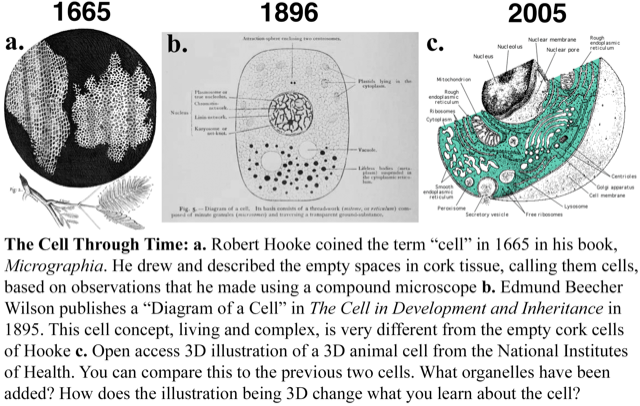
Image sources: (a) Hooke, Robert. Micrographia: or Some Physiological Descriptions of Minute Bodies Made by Magnifying Glasses. With Observations and Inquiries Thereupon. The Royal Society, 1665. (b) Wilson, Edmund Beecher. The Cell in Development and Inheritance. London: Macmillan and Co., 1896. http://dx.doi.org/10.5962/bhl.title.6239 (c) National Institutes of Health. "Diagram of an Animal Cell in Three Dimensions.” Wikimedia Commons, 2005.