Opening the Black Box of Late Neurodevelopment
WOODS HOLE, Mass. – The nematode worm, C. elegans, is one of the most widely used animal models in biological research. In 1998, it became the first animal genome to be fully sequenced and, since then, expansive research has been done to understand the function, expression, and interaction of its genes. Nearly every stage of C. elegans development and life has been studied in great detail – except the final six hours of embryogenesis.
Now, a new study emerging from a collaboration at the Marine Biological Laboratory (MBL) zeroes in on that critical time of development, by deploying innovative imaging techniques and computational tools. “There's a lot of interesting neurodevelopment that's going on during that period, so we wanted to capture that using all the strengths of C. elegans,” says Evan Ardiel, research fellow at Massachusetts General Hospital and Harvard Medical School and co-first author of the study, recently published in eLife.
The interdisciplinary team sought to better understand how the behavior of the embryo changes as the nervous system is coming online. Their paper provides the first detailed look at those embryonic behaviors and their progression during neurodevelopment.
“There's some reason to think that the earliest behaviors and the earliest circuits may have an outsized role overall in neurodevelopment and that what happens earliest on can have enduring effects on the developmental outcome at the individual,” says co-author Joshua Kaplan, professor at Harvard Medical School and Massachusetts General Hospital.
The team discovered that early on, the embryo repeatedly flips over and then slows down its motion before showing mature, adult-like motion in the last phase of embryogenesis. During the phase right before hatching, embryos begin crawling, moving forward and backward, and, importantly, going through rhythmic patterns of rest. The authors termed this sleep-like behavior “slow wave twitch (SWT)”.
This is the first paper to fully map out the movements happening in the last few hours of embryonic development in C. elegans and show a rhythmic, sleep-like state within that time period. Going forward, the team hopes to understand the function of SWT in aiding either development or neural circuit functioning.
Imaging Challenges and Solutions
C. elegans embryos are very delicate and sensitive to light, which creates a challenge for microscopy. When put under too much light, they arrest and stop developing, so many scientists, including senior author and MBL Fellow Hari Shroff, have adopted and developed light-sheet fluorescence microscopes (LSFM) that can image samples rapidly and gently while still capturing the whole 3D sample.
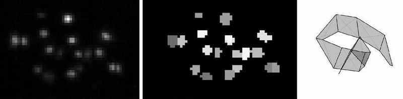
While LSFM and fluorescent markers on the sides of the developing animal, in principle, allow scientists to monitor its posture as it grows, tracking various parts of the animal during rapid embryo movement was still a challenge.
Andrew Lauziere, co-first author, NIH Intramural Research Fellow and graduate student at University of Maryland, used the different points along what he calls a “little motion capture suit” on the embryo to build improved tracking software. Some parts of the animal move together, an assumption that allowed the modeling of movement to be more accurate than assuming independent movement. Still challenging to model are the animal’s fast jerks, which the imaging rate is not quite quick enough to capture, but the new computational tool Lauziere developed allows scientists to visualize and manually correct errors from automated tracking.
“One new technical contribution here was the development of a tracking framework that simultaneously accounted for correlated embryo movements, incorporated the animal anatomy, and permitted manual correction of errors caused by embryo jerking movements,” says Shroff, senior investigator at NIH at the time of the work and currently Senior Group Leader at Janelia Research Campus.

Connecting Neural Activity and Behavior
After using this technique to create a library of all the embryo’s 3D postures, each of which were precisely temporally marked, the team then used those postures to characterize movements within the embryo as it develops, including the slow-wave twitch phase before hatching.
By studying a C. elegans mutant, the team discovered that the last phase of embryonic movement, containing SWT, depended on neuronal activity, whereas the earlier two phases did not. “Maybe it's not surprising to the C. elegans folks, but I always find it somewhat amazing,” says Shroff, that the embryos’ early movements don’t require neuronal input.
Because it was already known that larvae and adults rely on two specific neurons to go through restful states, the team investigated whether mutant embryos that lack one of these two neurons might still show SWT behaviors in the last phase of embryogenesis. They found that SWT depends on one of the neurons (RIS) to release a specific neurotransmitter and without it, SWT doesn’t occur. So SWT seems to share neuronal circuitry with sleep in larvae and adult C. elegans.
The team plans to continue much of their collaborative work at MBL, especially when it comes to testing out new microscopes and imaging tools. “I’m not a microscope geek. I’m not a computational biologist. Yet we all worked pretty fluently with each other,” reflects Kaplan. “And that’s a bit of the magic of Woods Hole; It brings really interdisciplinary efforts to the front and it propagates that in new students every year.”
Citation:
Evan L Ardiel, Andrew Lauziere, Stephen Xu, Brandon J Harvey, Ryan Patrick Christensen, Stephen Nurrish, Joshua M Kaplan, Hari Shroff (2022) Stereotyped behavioral maturation and rhythmic quiescence in C.elegans embryos eLife 11:e76836 doi.org/10.7554/eLife.76836
—###—
The Marine Biological Laboratory (MBL) is dedicated to scientific discovery – exploring fundamental biology, understanding marine biodiversity and the environment, and informing the human condition through research and education. Founded in Woods Hole, Massachusetts in 1888, the MBL is a private, nonprofit institution and an affiliate of the University of Chicago.