Shinya Inoué, Pioneer in Microscopy and Imaging of Live Cells, Dies at 98

WOODS HOLE, Mass.—With great sadness, the Marine Biological Laboratory (MBL) announces the passing of Shinya Inoué, Ph.D., former MBL Trustee and pioneering innovator in microscopy and live-cell imaging, on Sept. 30 in Falmouth, Mass. He was 98 years old.
A Celebration of Shinya Inoué’s Life will be held at the MBL on October 26, 2019 at the MBL Club, 100 Water Street, Woods Hole from 12 – 2 PM. Refreshments and light food will be available from 12 – 1 PM followed by a Celebration of Dr. Inoué’s life from 1 -2 PM. Friends, families, and colleagues are encouraged to share their stories and remembrances. A memorial notice provided by the Inoué family is here.
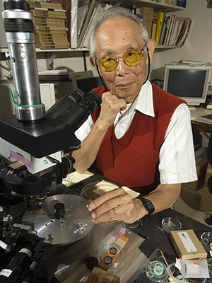 Shinya Inoué at the MBL in 2006. Credit: Tom Kleindinst
An extraordinary scientific leader and mentor at the MBL for more than half a century, Inoué was named Distinguished Scientist, the laboratory’s highest honor, in 1986. One of Inoué’s greatest contributions was to introduce the era of live-cell imaging by using polarized light microscopy to explore the intricacies of cellular structure and dynamics.
Inoué’s dual careers as a microscopist and cell biophysicist were elegantly complementary: Each refinement of his hand-built microscopes led to a clearer understanding of fundamental life processes, including cell division, fertilization, and early embryonic development. Disorder of these cellular events can cause critical diseases and growth abnormalities.
Inoué was also the co-inventor of video microscopy, a revolutionary advance that ushered in the modern era of electronic imaging, which quickly advanced to digital imaging. Important advances in video microscopy were developed at the MBL in the early 1980s.
Inoué’s far-reaching impact at the MBL began in the 1950s when he was a visiting investigator from Princeton University and continued after he joined the MBL as a senior scientist in 1982. He founded the Architectural Dynamics in Living Cells Program in 1992, an international, collaborative center for innovation in light microscopy that continues today under the umbrella of the MBL’s Bell Center.
As founding director of the MBL Analytical and Quantitative Light Microscopy course in 1979, Inoué pioneered an innovative commercial/academic course format that still today provides a fertile ground for beneficial interactions between the microscopy industry and the academic research community. This collaborative format has become standard, adopted by microscopy courses the world over.
With his son, Ted Inoué, Shinya co-founded Universal Imaging Corporation in 1984, one of the first digital image analysis and electronic microscope control software companies. This company produced the first digital image analysis computer software, Image-1, which was eventually renamed MetaMorph and set the standard for all image analysis software made today.
Inoué served on the MBL Board of Trustees from 1970 to 1977, 1979 to 1986, and 1992 to 1996 and was a member of the MBL Society from 1962 to 2012. He and his wife, Sylvia, established several funds and endowed lectureships at the MBL to support research and training in microscopy. He also served on the faculty of the MBL’s Physiology course and Optical Microscopy course for many years.
“For the last 30 years, I had the great fortune to work with Shinya Inoué, who was an exacting and demanding, yet patient and always generous mentor, who taught by example, combining a passion both for creating tools and applying them to reveal the mysteries of life,” said MBL Senior Scientist Rudolf Oldenbourg. “Inoué not only was an outstanding scientist, but he is universally respected for his kind and thoughtful ways, for his humanity, and his attention to personal relationships.”
“We have a lost a giant in cell biology and a wonderful human being," said colleague Ron Vale of University of California, San Francisco. "Shinya was the pioneer in studying the dynamics of living cells by microscopy, an approach that is widespread today. He developed many new methods in microscopy, and his observations led to many unique insights in the working of cells. Shinya left behind a lasting legacy of contributions in cell biology. He will be widely missed.”
The son of a Japanese diplomat, Inoué was born in 1921 in London, England. His distinguished career began in the 1940s at Tokyo University, where as an undergraduate he studied with cell biologist and MBL summer investigator Katsuma Dan. Dan presented Inoué with a challenge: Build a microscope that will allow us to see the mitotic spindle—the transient structure that moves chromosomes in the dividing cell—in a living sea-urchin egg. While the great microscopists of the 19th and early 20th centuries had produced fine images of cellular parts, including the mitotic spindle, these were generally produced in fixed (nonliving) cells. Inoué rose to Dan’s challenge, building his first polarized light microscope out of various found parts—including a discarded machine-gun base and a tin tea can—at Misaki Marine Biological Station in 1947, in the aftermath of World War II.
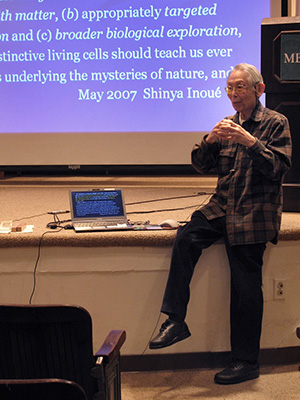 Shinya Inoué teaching in the MBL Physiology course in 2007. Credit: Dyche Mullins
With the assistance of Katsuma Dan’s wife, biologist Jean Clark Dan, Inoué entered Princeton University in 1948, where he was mentored by cytologist Kenneth W. Cooper and graduated with a Ph.D. in biology in 1951. While at Princeton, Inoué improved his polarized light microscope (now nicknamed the “Shinya Scope”) and in 1951 he used it to prove the universal existence of the spindle fibers, the dynamic protein filaments that move chromosomes in the dividing cell. Inoué announced this landmark discovery in the MBL’s Lillie Auditorium, where he premiered a movie of dividing cells that clearly showed the action of the spindle fibers in the mitotic spindle. It was the first major accomplishment in a career devoted to delving into the mysteries of living cells.
“In an attempt to better understand how cells divide, Dr. Inoué made a series of epochal innovations in the development of light microscopy,” said Akihito, the former Emperor of Japan, in awarding Inoué the 2003 International Prize for Biology. “These advances rendered it possible to directly observe dynamic changes in the supramolecular structure of living cells during cell division. This contributed immensely to advancing research in such fields of cell division, cytoskeleton, and cell motility … The products of Dr. Inoué’s research are widely utilized by researchers around the world and contribute immensely to the advancement of biological sciences.”
Shortly after joining the MBL in 1982, Inoué and Robert and Nina Allen independently discovered at the MBL that using a video camera (rather than the eye) to record images from a microscope brought great gains in image clarity. Inoué combined video microscopy with computer-assisted contrast enhancement, allowing one to see fine details of cells that had never been seen before. It was the birth of the electronic era in microscopy.
Over five decades, Inoué built seven generations of his Shinya Scope, with technical improvements each time, and in the late 1990s he invented the centrifuge polarizing microscope. Inoué held four U.S. patents for his microscopes and authored more than 100 scientific papers, many of which are collected in The Collected Works of Shinya Inoué: Microscopes, Living Cells, and Dynamic Molecules (2008: World Scientific Publishing Co.) He also authored the book Video Microscopy (1986: Plenum Press).
Prior to his move to the MBL, Inoué was professor of biology at University of Pennsylvania (1966-1982), where he directed the Program in Biophysical Cytology; professor and department chair of cytology at Dartmouth Medical School (1959-1966); associate professor at University of Rochester (1954-1959); assistant professor at Tokyo Metropolitan University (1953-1954); and instructor at University of Washington (1951-1953).
Inoué was the recipient of numerous awards and honors. They include the Order of the Sacred Treasure, Gold Rays with Neck Ribbon Award from the Government of Japan (2010); the International Prize for Biology from the Japan Society for the Promotion of Science (2003); membership in the National Academy of Sciences and the American Academy of Arts and Sciences; the Distinguished Scientist Award from the Microscopy Society of America (1995); and the E.B. Wilson Award from the American Society for Cell Biology (1992). He was honored to lecture on and demonstrate his work to Japan’s Emperor Showa in 1947, 1953, and 1975.
He leaves his wife, Sylvia (McCandless) Inoué, five children, six grandchildren, and one great-grandchild.
Other Memorial Notices and Resources:
Video: Shinya Inoué Celebration of Life event at MBL, Oct. 26, 2019
Reminiscences of Shinya Inoué by colleagues (collected by Rudolf Oldenbourg and Tomami Tani)
Shinya Inoué Obituary in Cell (by Timothy Mitchison)
Shinya Inoué Obituary in The Biological Bulletin (by Lisa A. Cameron)
Exhibit on Shinya Inoué prepared by the History of the Marine Biological Laboratory Project
In Memory of Shinya Inoue, American Society for Cell Biology (by Greenfield Sluder)
—###—
The Marine Biological Laboratory (MBL) is dedicated to scientific discovery – exploring fundamental biology, understanding marine biodiversity and the environment, and informing the human condition through research and education. Founded in Woods Hole, Massachusetts in 1888, the MBL is a private, nonprofit institution and an affiliate of the University of Chicago.