The Winners of the 2019 MBL Photo Contest

The Marine Biological Laboratory is excited to announce the winners of the 2019 MBL Photo Contest. We received dozens of stunning images of life science specimens and macro photos highlighting MBL research. Below are the 12 photos chosen by our judges.
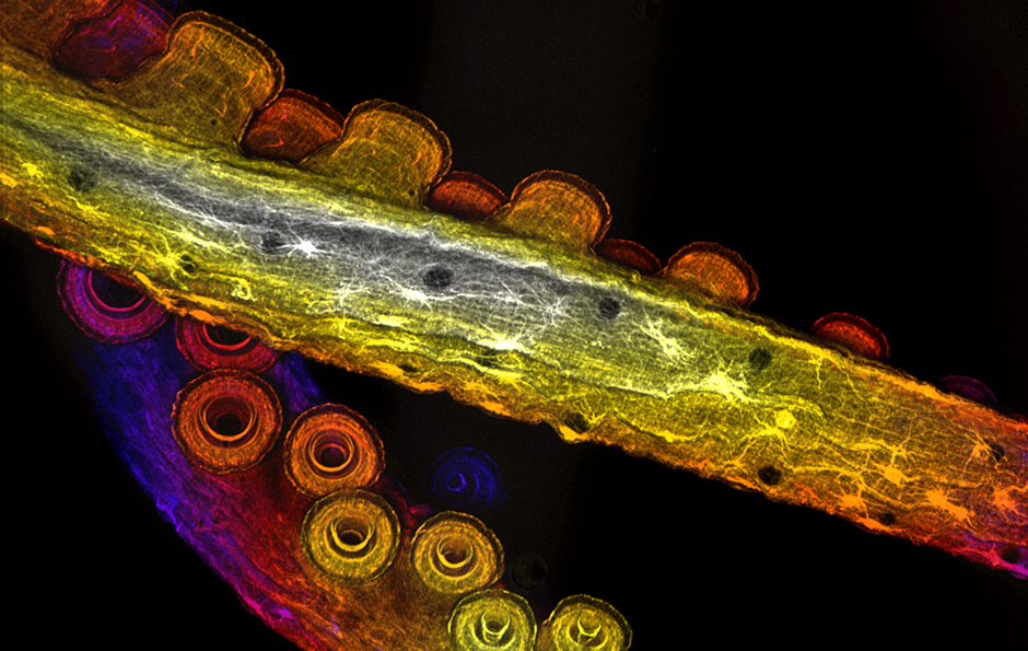 Cover Photo Winner Arm muscles of Octopus bimaculoides Allan Carrillo-Baltodano, Student
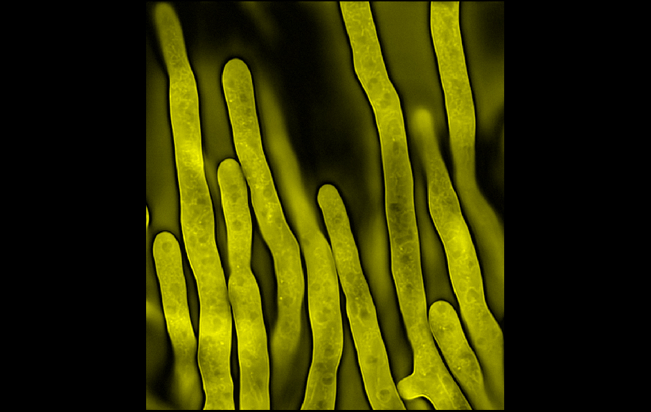 Filamentous fungus Ashbya gossypii Michael Shribak, Associate Scientist
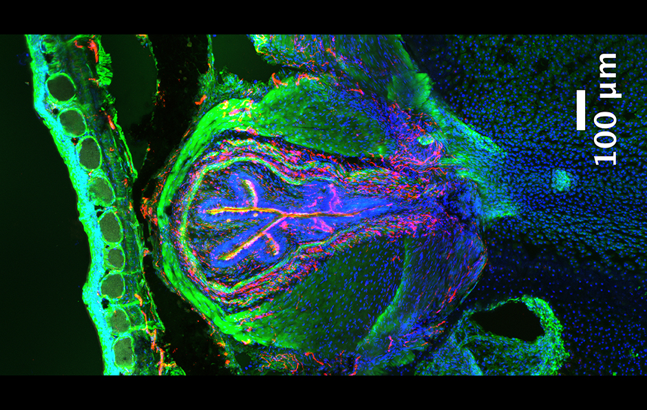 A transverse cross-section across the frog urostyle Gayani Senevirathne, Whitman Center Graduate Student, University of Chicago
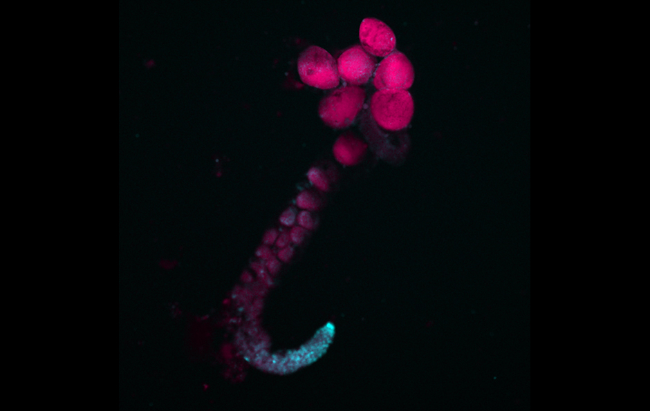 Ovarium of the ostracod Heterocypris incongruens, showing the presence of endosymbiontic bacteria Isabelle Schön, Whitman Center Fellow
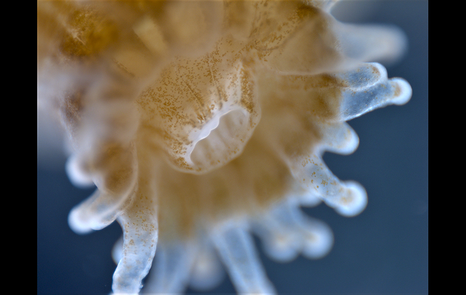 Northern Coral Cup Mayra Sánchez García, Research Assistant II
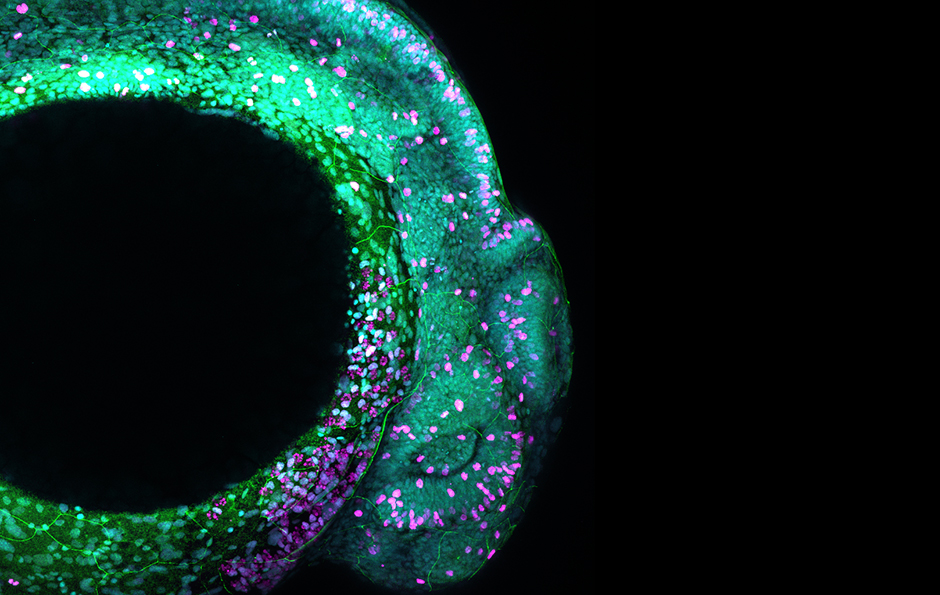 Zebrafish Embryo Arnon Jurberg, Assistant Professor, University Estácio de Sá, Brazil
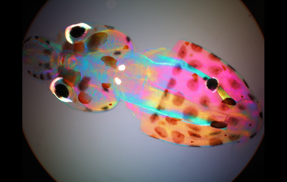 Birefringence of Doryteuthis pealeii hatchling Elizabeth Lee, Whitman Center Assistant, University of Chicago
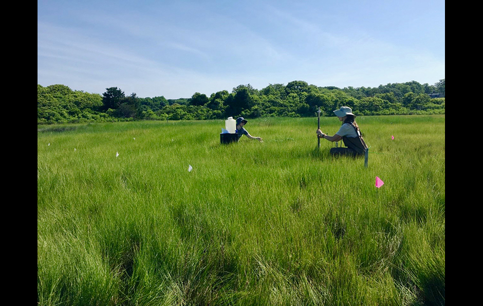 Elevation in Great Sippewissett Salt Marsh Kelsey Chenoweth, Research Assistant I, Marine Biological Laboratory
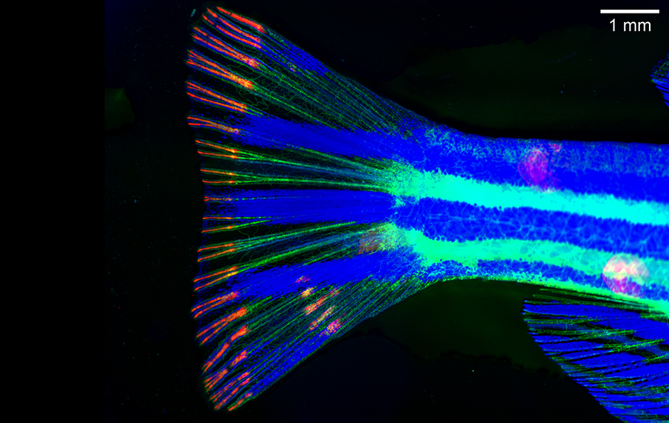 Resect, Regrow and Regenerate Victoria Deneke, Whitman Center Assistant Research Institute for Molecular Pathology, Austria
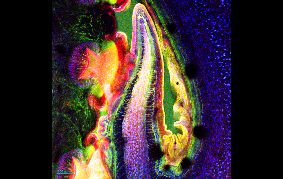 Octopus arm, stained with vital dyes Stephen Senft, Research Associate, Marine Biological Laboratory
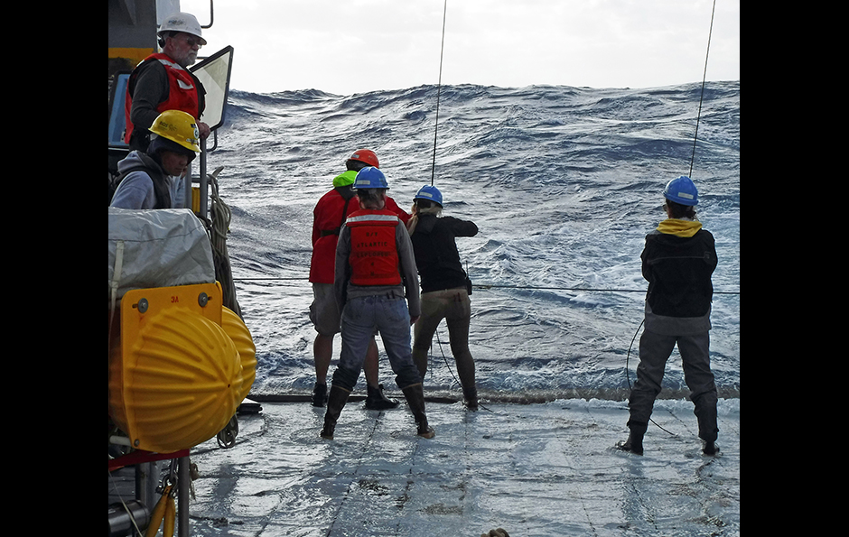 Oceanographic sampling in rough weather JC Weber, Senior Research Assistant, Marine Biological Laboratory
Further image information:
Arm muscles of Octopus bimaculoides
Description: Detail of the muscles in the arms of Octopus bimaculoides, color coded for depth.
Photographer: Allan Carrillo-Baltodano, Student, Queen Mary University of London
Microscope: ZEISS Axio Zoom.V16
Camera: ZEISS Axiocam
Filamentous fungus Ashbya gossypii
Description: Image of filamentous fungus Ashbya gossypii, made in cooperation with MBL Fellow Amy Gladfelter, University of North Carolina, Chapel Hill.
Photographer: Michael Shribak, Associate Scientist, Marine Biological Laboratory
Microscope: Olympus BX61
Camera: Lumenera Infinity 3-1M
A transverse cross-section across the frog urostyle
Description: Neuronal distribution within the urostyle taken from OCT-frozen samples and stained for three different antibodies: extracellular matrix (Phalloin - green), neurons (acetylated tubulin - red), and nuclei (DAPI - blue).
Photographer: Gayani Senevirathne, Whitman Center Graduate Student, University of Chicago
Microscope: ZEISS LSM 880
Camera: Airyscan
Ostracod, Heterocypris incongruens, ovarium with endosymbiontic bacteria
Description: Ovarium in initial stage (bottom) with oocyte formation and unfertilized parthenogenetic eggs (top). Yolk tissue (red), bacteria (blue). Produced with Jessica Mark Welch, MBL and Koen Martens, Royal Belgian Institute of Natural Sciences.
Photographer: Isabelle Schön, Whitman Center Fellow, Royal Belgian Institute of Natural Sciences
Microscope: ZEISS LSM 780
Camera: GaAsP spectral detector
Northern Coral Cup
Description: A close-up image of a polyp of the Astrangia poculata coral colony
Photographer: Mayra Sánchez García, Research Assistant II, Marine Biological Laboratory
Microscope: ZEISS Axio Zoom V16
Camera: ZEISS Axiocam 503 color
Baby Bimac
Description: Hatchling Octopus bimaculoides raised in the MBL’s Marine Resources Center and used to extract DNA and RNA for genomics analysis.
Photographer: Kirsten Sadler Edepli, Whitman Center Associate, New York University Abu Dhabi
Camera: iPhone 8
Zebrafish Embryo
Description: Head of a zebrafish embryo, showing the eye and brain (right). In magenta, proliferating cells.
Photographer: Arnon Jurberg, Assistant Professor, University Estácio de Sá, Brazil
Microscope: ZEISS LSM 800
Camera: PMTs
Birefringence of Doryteuthis pealeii hatchling
Description: Squid hatchling observed through a polychromatic polscope, where ordered structures such as muscle are colored.
Photographer: Elizabeth Lee, Whitman Center Assistant, University of Chicago
Microscope: Olympus IX81inverted microscope
Camera: Olympus DP73 color CCD camera
Elevation in Great Sippewissett Salt Marsh
Description: Students using a water-leveling method to measure elevation in Great Sippewissett Marsh (Falmouth, MA) plots which have been studied by MBL scientists for nearly 50 years.
Photographer: Kelsey Chenoweth, Research Assistant I, Marine Biological Laboratory
Camera: iPhone 6s
Resect, Regrow and Regenerate
Description: Regenerating zebrafish fin 7 days post amputation. Osteblasts are labeled in red, vasculature in green, and bright field image is overlayed in blue.
Photographer: Victoria Deneke, Whitman Center Assistant, Research Institute for Molecular Pathology, Austria
Camera: Leica
Octopus arm, stained with vital dyes
Description: Coiled distal arm of a young cephalopod, Octopus bimaculoides, labeled in vivo using multiple dyes.
Photographer: Stephen Senft, Research Associate, Marine Biological Laboratory
Microscope: ZEISS LSM 780
Camera: Laser scanning microscope
Oceanographic sampling in rough weather
Description: MBL researchers deploy an in situ pump to collect deep ocean particles in rough seas off Bermuda.
Photographer: JC Weber, Senior Research Assistant, Marine Biological Laboratory
Camera: Fujifilm XP90