Full Name
Zak Swartz
Title
Assistant Scientist

- Email:
- Phone:
- Fax:
- CV:
- ORCID ID:
0000-0002-3264-6880
B.S., Developmental Biology, University of Rochester, 2007
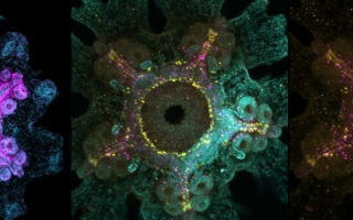
We are fascinated by how animals make eggs, and how eggs make animals. Our laboratory is defining molecular mechanisms that underly the development of an egg from its precursor cell, the oocyte. To address these questions, we are working at the interface between cell biology and development, with the sea star Patiria miniata and its relatives. Sea stars produce millions of oocytes throughout their lives, and are amenable to an extensive range of molecular and genetic tools, as well as high resolution light microscopy. As relatives to the animal group that includes vertebrates like ourselves, the sea star can teach us important lessons relevant for human reproductive health, disease, and aging. Current projects in the lab include:

Tuning cell division for reproduction and in response to a changing ocean
Oocytes must undergo an acrobatic series of divisions during a specialized cell cycle called meiosis, which is shared amongst nearly all animals. They must also be fertilized to incorporate the male genome, and then switch to mitosis, which divides the egg into individual embryonic cells. These divisions must occur precisely, as errors can lead to loss of fertility and developmental disorders. We are asking how phosphorylation and regulated translation coordinate these rapid changes in division strategy. Incredibly, in sea stars and other ecologically important animals, these events all take place exposed in the open ocean. Therefore, a major goal of our group is to determine how cellular processes withstand fluctuations within the natural habitat.

Development of oocytes within the ovarian environment
Oocytes do not arise in isolation, but rather surrounded by specialized cells within the ovary. Oocytes communicate biochemically with these surrounding cells, but the extent of these pathways are not fully defined. Unlike mammals, sea stars display a tremendous reproductive longevity, and continue to produce new oocytes throughout their lifespan. This unique biology, along with an in vitro culture system, will allow us to define the cellular organization of the ovary, and the ways in which developing oocytes communicate with surrounding cells. This work will help us to uncover conserved processes in oogenesis important for fertility and the evolution of the ovary.
Establishment of organismal body axes
In many animals, the primary body axis (e.g. heads vs. tails, or anterior vs. posterior), is programmed directly into the egg. What are the molecular cues that define this axis and how do they transmit that information into the developing embryo? We are determining how an ancient regulator of anterior-posterior identity, a protein called Dishevelled, becomes specifically activated at the bottom portion of the oocyte that will give rise to the posterior embryo. This work will help us learn more about how the animal body plan is established, as well as its evolution.


Swartz SZ, Tan TH, Perillo M, Fakhri N, Wessel GM, Wikramanayake AH, Cheeseman IM. Polarized Dishevelled dissolution and reassembly drives embryonic axis specification in sea star oocytes. Curr Biol. 2021;31(24):5633-41 e4. Epub 2021/11/06. doi: 10.1016/j.cub.2021.10.022.
Swartz SZ, Nguyen HT, McEwan BC, Adamo ME, Cheeseman IM, Kettenbach AN. Selective dephosphorylation by PP2A-B55 directs the meiosis I-meiosis II transition in oocytes. Elife. 2021;10. Epub 2021/08/04. doi: 10.7554/eLife.70588.
Wigbers MC, Tan TH, Brauns F, Liu J, Swartz SZ, Frey E, Fakhri N. A hierarchy of protein patterns robustly decodes cell shape information. Nature Physics. 2021;17(5):578-84. doi: 10.1038/s41567-021-01164-9
Swartz SZ, McKay LS, Su KC, Bury L, Padeganeh A, Maddox PS, Knouse KA, Cheeseman IM. Quiescent Cells Actively Replenish CENP-A Nucleosomes to Maintain Centromere Identity and Proliferative Potential. Dev Cell. 2019;51(1):35-48 e7. Epub 2019/08/20. doi: 10.1016/j.devcel.2019.07.016.